Question
Draw illustrations to bring out the anatomical difference between
(a) Monocot root and Dicot root
(b) Monocot stem and Dicot stem
(a) Monocot root and Dicot root
(b) Monocot stem and Dicot stem
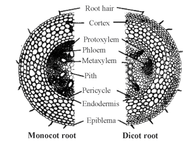
| Features | Monocot root | Dicot root |
| (i) Cortex | Comparatively narrow. | Very wide. |
| (ii) Endodermis | Less thickened and casparian strips are more prominent. | Later become highly thickened. Casparian strips are visible only in young root. |
| (iii) Passage cells | Generally absent. | Generally occur opposite the proto-xylem point. |
| (iv) Pericycle | Produces lateral roots, cork cambium and part of the vascular cambium. | Produces lateral roots only. |
| (v) Vascular bundles | 2 to 5 or some-times 8. | 8 or more in number. |
| (vi) Pith | Either absent or very small | Well-developed. |
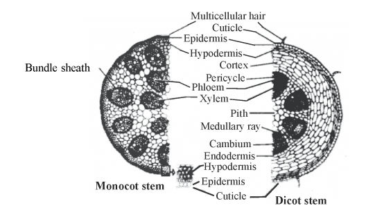
| Features | Monocotyledonous stem | Dicotyledonous stem |
| (i) Vascular bundles | (a) Scattered (b) Conjoint, collateral, closed. (c) Bundle sheath usually present. (d) Phloem parenchyma absent. (e) Xylem vessels arranged either in Y or V shaped manner. | (a) Vascular bundles in ring (b) Conjoint, collateral or bicollateral and open. (c) Bundle sheath absent. (d) Phloem parenchyma present. (e) Not so. |
| (ii) Pith (Medulla) | Absent | Made up of parenchymatous cells situated in the centre of stem. |
| (iii) Ground tissue | Ground tissue is not differentiated into the cortex and pith. | Differentiated into the cortex and pith. |
| (iv) Hypodermis | Usually sclerenchymatous | Collenchymatous. |
| (v) Endodermis | Absent | One layered, starchy sheath which is usually not well differentiated. |
| (vi) Pericycle | Absent | Made up of one or more layers of parenchymatous and/or sclerenchymatous cells. |
| (vii) Medullary rays | Absent | Found in between vascular bundles. |
Generate a complete, print-ready paper with questions like this in minutes — across 16+ boards, with answer keys.