Question
Draw labelled diagrams
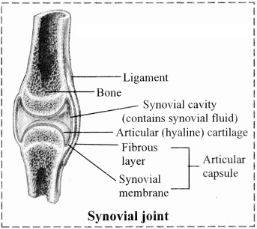
Synovial joint.
Synovial joint.
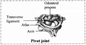
1. Pivot joint: In this type of joint, the rounded or pointed surface of one bone articulates with a ring formed partly by another bone and partly by the ligament. Rotation only around its own longitudinal axis is possible. e.g. in joint between atlas and axis vertebrae, head turns side ways to form ‘NO’ joint.
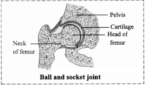
2. Ball and socket joint: The ball like surface of one bone fits into cup like depression of another bone forming a movable joint. Multi-axial movements are possible. This type of joint allows movements along all three axes and in all directions. e.g. Shoulder and hip joint.
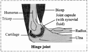
3. Hinge joint: In a hinge joint, convex surface of one bone fits into concave surface of another bone. In most hinge joints one bone remains stationary and other moves. The angular opening and closing motion (like hinge) is possible. In this joint only mono-axial movement takes place like flexion and extension. e.g. Elbow and knee joint.
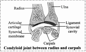
4. Condyloid joint: It is an ellipsoid joint. The convex oval shaped projection of one bone fits into oval shaped depression in another bone. it is a biaxial joint because it permits movement along two axes viz, flexion, extension, abduction, adduction and circumduction is possible. e.g. Metacarpophalangeal joint.
5. Gliding joint: It is a planar joint, where the articulating surfaces of bones are flat or slightly curved. These joints are non-axial because the motion they allow does not occur along an axis or a plane. e.g. Intercarpal and intertarsal joints.
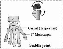
6. Saddle joint: This joint is a characteristic of Homo sapiens. Here the articular surface of one bone is saddle-shaped and that of other bone fits into saddle (each bone forming this joint have both concave and convex areas). It is a modified condyloid joint in which movement is somewhat more free. It is a biaxial joint that allows flexion, extension, abduction, adduction and circumduction.
e.g. Carpometacarpellar joint between carpal (trapezium) and metacarpal of thumb.
Generate a complete, print-ready paper with questions like this in minutes — across 16+ boards, with answer keys.