Gujarat BoardEnglish MediumSTD 11 ScienceBIOLOGYMorphology of Flowering Plants5 Marks
Question
With suitable diagrams explain the structures of dicot and monocot root.
✓
Answer
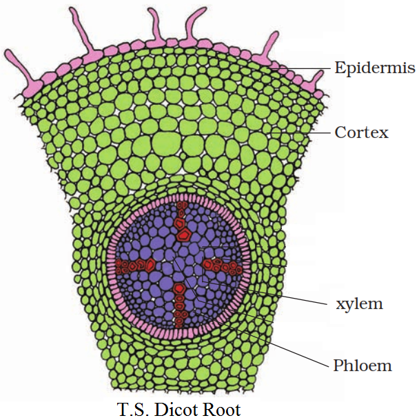
Dicotyledonous Root:
Epidermis: The outermost layer is epidermis. Many of the epidermal cells protrude in the form of unicellular root hairs.
Cortex: The cortex consists of several layers of thin-walled parenchyma cells with intercellular spaces.
Endodermis: The innermost layer of the cortex is called endodermis. It comprises a single layer of barrel-shaped cells without any intercellular spaces. The tangential as well as radial walls of the endodermal cells have a deposition of water be impermeable, waxy material suberin in the form of caspariun strips. Next to endodermis lies a few layers of thick-walled parenchyomatous cells referred to as pericycle. Initiation of lateral roots and vascular cambium during the secondary growth takes place in these cells. The pith is small or inconspicuous. The parenchymatous cells which lie between the xylem and the phloem are called conjuctive tissue. There are usually two to four xylem and phloem patches. Later, a cambium ring develops between the xylem and phloem. All tissues on the innerside of the endodermis such as pericycle, vascular bundles and pith consuute the stele.
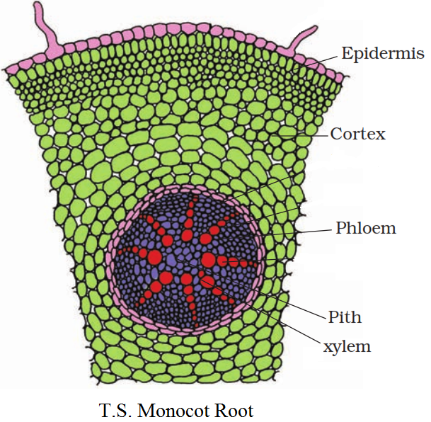
Monocotyledonous Root: The anatomy of the monocot root is similar to the dicot root in many respects. It has epidermis, cortex, endodermis, pericycle, vascular bundles and pith. As compared to the dicot root which have fewer xylem bundles, there are usually more than six (polyarch) xylem bundles in the monocot root. Pith is large and well developed. Monocotyledonous roots do not undergo any secondary growth.
Need a full question paper?
Generate a complete, print-ready paper with questions like this in minutes — across 16+ boards, with answer keys.