Question 15 Marks
How does cytokinesis in plant cells differ from that in animal cells?
Answer
View full question & answer→Differences between plant cytokinesis and animal cytokinesis
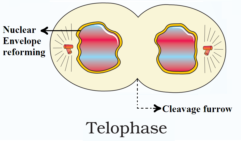
| Plant Cytokinesis | Animal Cytokinesis | ||
| i. | It occurs by cell plate formation. | i. | It occurs by cleavage. |
| ii. | The cell plate appears at the centre and extends outwards. | ii. | Cleavage begins at the periphery and proceeds inwards |
| iii. | Fusion of vesicles begins cell plate formation. | iii. | Cleavage is started by contraction of a peripheral ring of microfilaments. |
| iv. | A midbody is not formed. | iv. | A midbody of dense material is formed at the middle of the cell. |
 Which of these structures is responsible for,
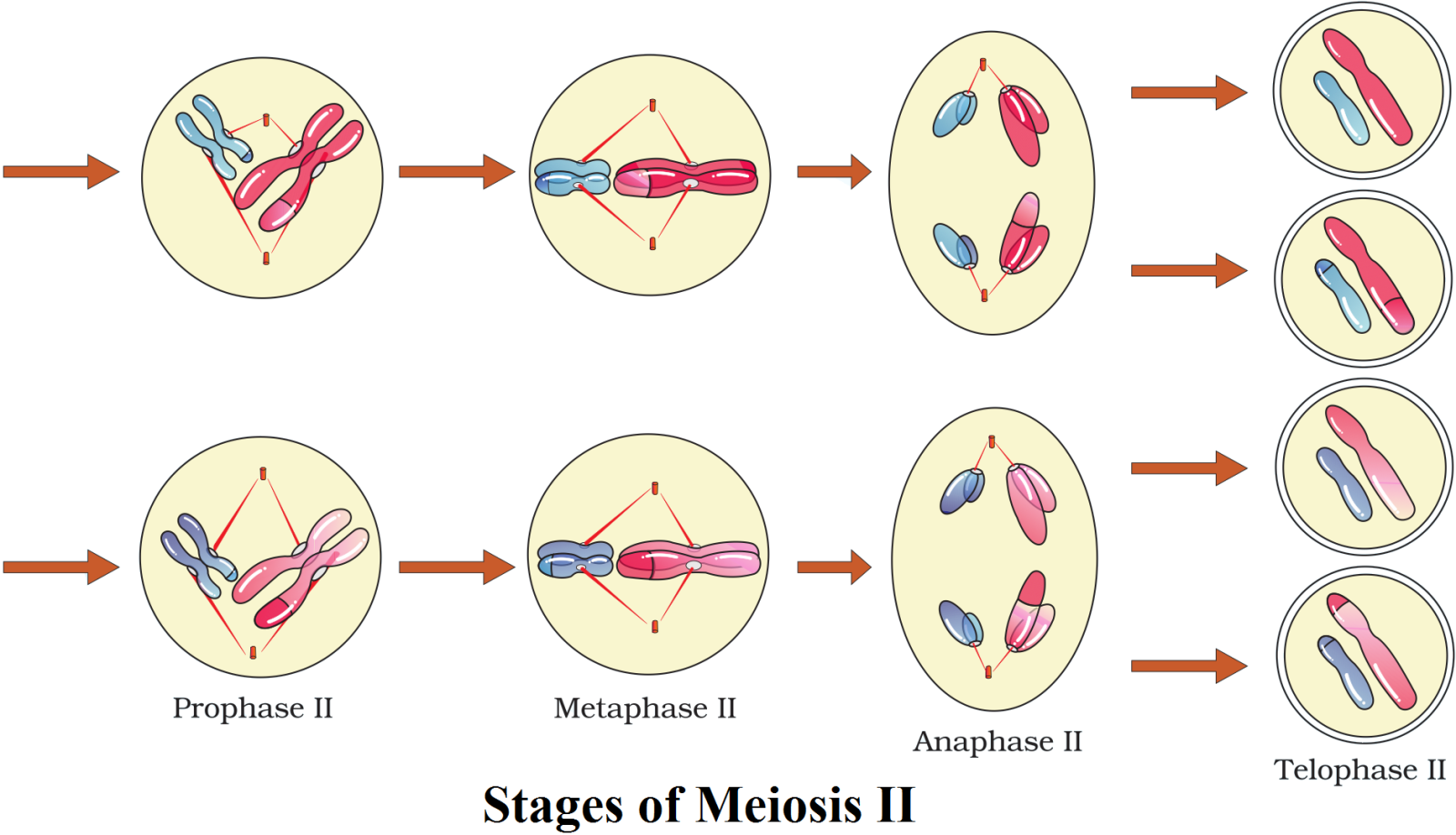
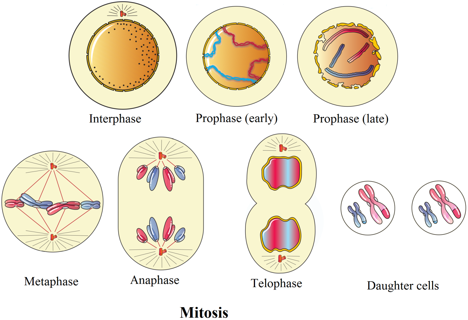
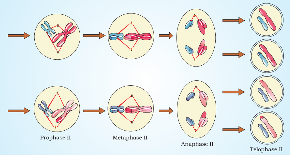 Meiosis II − Resembles Mitosis