Question 15 Marks
With suitable diagram describe animal cell.
Answer
View full question & answer→An animal cell has the following cell structures:
• Plasma membrane
• Endoplasmic reticulum
• Mitochondria
• Golgi body
• Lysosomes
• Ribosomes
• Vacuoles
• Nucleus
• Centriole
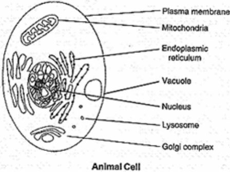
i. Plasma Membrane: This is also known as cell membrane. Plasma membrane is made up of lipid and protein. It is semi - permeable in nature. Certain substances are transported through plasma membrane by passive transport. Some substances get transported by osmosis and some by active transport. Active transport involves use of some carrier to facilitate transport. Apart from transport of materials, plasma membrane gives a shape and size to the animal cell.
ii. Endoplasmic Reticulum: These are networks of fine tubules extending from plasma membrane to nucleus. They work like pipelines and facilitate transport of substances from outside the cell to nucleus and cytoplasm. Depending on presence or absence of ribosomes ER can be either rough or smooth.
iii. Golgi Body: This is composed of many sack like structures stacked one over another. The function of golgi body is to package different materials, like carbohydrate, protein and lipid.
iv. Lysosome: Lysosome is a small spherical structure filled with digestive enzymes. The digestive enzyme helps in digesting foreign materials and waste products. Sometimes the lysosome digests the contents of cytoplasm which in turn kills the cell itself. That is why lysosome is also known as 'suicide bag of the cell'.
v. Ribosome: Ribosomes are small dot-like structures. They are made of two subunits. The function of the ribosome is to synthesize protein.
vi. Vacuoles: These are small fluid-filled structures. Vacuoles help in maintaining osmotic pressure inside the cell.
vii. Mitochondria: Mitochondria is a double membrane structure. The inner membrane is projected in finger-like structures, called cristae. The presence of cristae helps in increasing the inner surface are of mitochondria. Aerobic respiration takes place in the mitochondria and energy released is stored in the form of ATP (Adenosine triphosphate).
viii. Nucleus: Nucleus is covered by a nuclear membrane. The nucleus contains chromosomes which are genetic materials. Nucleus also controls various functions of the cell.
ix. Centriole: These are spindle-like structures. During cell division, they form spindle fibres.
• Plasma membrane
• Endoplasmic reticulum
• Mitochondria
• Golgi body
• Lysosomes
• Ribosomes
• Vacuoles
• Nucleus
• Centriole
i. Plasma Membrane: This is also known as cell membrane. Plasma membrane is made up of lipid and protein. It is semi - permeable in nature. Certain substances are transported through plasma membrane by passive transport. Some substances get transported by osmosis and some by active transport. Active transport involves use of some carrier to facilitate transport. Apart from transport of materials, plasma membrane gives a shape and size to the animal cell.
ii. Endoplasmic Reticulum: These are networks of fine tubules extending from plasma membrane to nucleus. They work like pipelines and facilitate transport of substances from outside the cell to nucleus and cytoplasm. Depending on presence or absence of ribosomes ER can be either rough or smooth.
iii. Golgi Body: This is composed of many sack like structures stacked one over another. The function of golgi body is to package different materials, like carbohydrate, protein and lipid.
iv. Lysosome: Lysosome is a small spherical structure filled with digestive enzymes. The digestive enzyme helps in digesting foreign materials and waste products. Sometimes the lysosome digests the contents of cytoplasm which in turn kills the cell itself. That is why lysosome is also known as 'suicide bag of the cell'.
v. Ribosome: Ribosomes are small dot-like structures. They are made of two subunits. The function of the ribosome is to synthesize protein.
vi. Vacuoles: These are small fluid-filled structures. Vacuoles help in maintaining osmotic pressure inside the cell.
vii. Mitochondria: Mitochondria is a double membrane structure. The inner membrane is projected in finger-like structures, called cristae. The presence of cristae helps in increasing the inner surface are of mitochondria. Aerobic respiration takes place in the mitochondria and energy released is stored in the form of ATP (Adenosine triphosphate).
viii. Nucleus: Nucleus is covered by a nuclear membrane. The nucleus contains chromosomes which are genetic materials. Nucleus also controls various functions of the cell.
ix. Centriole: These are spindle-like structures. During cell division, they form spindle fibres.