Question 14 Marks
Describe pregnancy and embryonic development.
Answer
View full question & answer→→ Immediately after implantation, the inner cell mass (embryo) differentiates into an outer layer called ectoderm and an inner layer called endoderm.
→ A mesoderm soon appears between the ectoderm and the endoderm.
→ These three layers give rise to all tissues (organs) in adults.
→ The inner cell mass contains certain cells called stem cells which have the potency to give rise to all the tissues and organs.
→ The human pregnancy lasts 9 months.
→ In human beings, after one month of pregnancy, the embryo's heart is formed. The first sign of growing foetus may be noticed by listening to the heart sound carefully through the stethoscope.
→ By the end of the second month of pregnancy, the foetus develops limbs and digits.
→ By the end of 12 weeks (first trimester), most of the major organ systems are formed, for example, the limbs and external genital organs are well-developed.
→ The first movements of the foetus and appearance of hair on the head are usually observed during the fifth month.
→ By the end of about 24 weeks (end of second trimester), the body is covered with fine hair, eye-lids separate, and eyelashes are formed.
→ By the end of nine months of pregnancy, the foetus is fully developed and is ready for delivery.
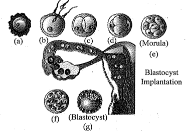
→ The zygote is formed at the ampulla-isthmus junction of the fallopian tube when the union of secondary oocyte and spermatozoa occurs. The zygote undergoes various divisions and changes before it reaches the uterus for implantation.
→ The figure shows the various stages of development of a zygote as it passes through the oviduct and uterus:
(a) The zygote divides into a two-celled stage wide via division or 1st cleavage while the ozygote is still in the isthmus of the oviduct.
(b) Several more mitotic divisions or cleavages occur in the 2 celled stage to form a 2, 4, 8 and finally a 16-celled stage of the zygote.The 16 celled stage is called the morula and various cells formed after cleavage are called blastomeres.
(c) Morula then changes into a blastocyst after a few more divisions and this stage contains a fluid filled cavity in the embryo. The blastomeres become arranged and line up into an outer layer of cells called the trophoblast and an inner mass of cells. The fluid filled cavity is called blastocoel.
(d) Implantation of the embryo occurs at this blastocyst stage by the help of trophoblast layer which embeds itself into the uterine endometrium.
→ A mesoderm soon appears between the ectoderm and the endoderm.
→ These three layers give rise to all tissues (organs) in adults.
→ The inner cell mass contains certain cells called stem cells which have the potency to give rise to all the tissues and organs.
→ The human pregnancy lasts 9 months.
→ In human beings, after one month of pregnancy, the embryo's heart is formed. The first sign of growing foetus may be noticed by listening to the heart sound carefully through the stethoscope.
→ By the end of the second month of pregnancy, the foetus develops limbs and digits.
→ By the end of 12 weeks (first trimester), most of the major organ systems are formed, for example, the limbs and external genital organs are well-developed.
→ The first movements of the foetus and appearance of hair on the head are usually observed during the fifth month.
→ By the end of about 24 weeks (end of second trimester), the body is covered with fine hair, eye-lids separate, and eyelashes are formed.
→ By the end of nine months of pregnancy, the foetus is fully developed and is ready for delivery.
→ The zygote is formed at the ampulla-isthmus junction of the fallopian tube when the union of secondary oocyte and spermatozoa occurs. The zygote undergoes various divisions and changes before it reaches the uterus for implantation.
→ The figure shows the various stages of development of a zygote as it passes through the oviduct and uterus:
(a) The zygote divides into a two-celled stage wide via division or 1st cleavage while the ozygote is still in the isthmus of the oviduct.
(b) Several more mitotic divisions or cleavages occur in the 2 celled stage to form a 2, 4, 8 and finally a 16-celled stage of the zygote.The 16 celled stage is called the morula and various cells formed after cleavage are called blastomeres.
(c) Morula then changes into a blastocyst after a few more divisions and this stage contains a fluid filled cavity in the embryo. The blastomeres become arranged and line up into an outer layer of cells called the trophoblast and an inner mass of cells. The fluid filled cavity is called blastocoel.
(d) Implantation of the embryo occurs at this blastocyst stage by the help of trophoblast layer which embeds itself into the uterine endometrium.