Question 15 Marks
List out the important features of Chromosomes.
Answer
View full question & answer→The four important features of the chromosome are:
1. The shape of the chromosome is specific: The long, thin, lengthy structured chromosome contains a short, constricted region called centromere. A centromere may occur any where along the chromosome, but it is always in the same position on any given chromosome. The number of chromosomes per species is fixed: For example the mouse has 40 chromosomes, the onion has 16 and humans have 46.
2. Chromosomes occur in pairs: The chromosomes of a cell occur in pairs, called homologous pairs. One of each pair come originally from each parent. Example, human has 46 chromosomes, 23 coming originally from each parent in the process of sexual reproduction. Chromosomes are copied: Between nuclear divisions, whilst the chromosomes are uncoiled and cannot be seen, each chromosome is copied. The two identical structures formed are called chromatids.
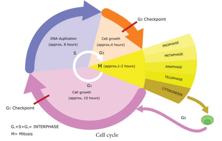 3. Nutrient deprivation: Lack of growth factors or density dependant inhibition. Undergo metabolic changes and enter into $G _0$ state. Biochemicals inside cells activates the cell division. The proteins called kinases and cyclins activate genes and their proteins to perform cell division. Cyclins act as major checkpoint which operates in $G _1$ to determine whether or not a cell divides.4. $G_0$ Phase: Some cells exit $G_1$ and enters a quiescent stage called $G_0$, where the cells remain metabolically active without proliferation. Cells can exist for long periods in $G_0$ phase. In $G_0$ cells cease growth with reduced rate of RNA and protein synthesis. The $G_0$ phase is not permanent. Mature neuron and skeletal muscle cell remain permanently in $G_0$. Many cells in animals remains in $G_0$ unless called on to proliferate by appropriate growth factors or other extracellular signals. $G_0$ cells are not dormant.5. S phase - Synthesis phase - cells with intermediate amounts of DNA. Growth of the cell continues as replication of DNA occur, protein molecules called histones are synthesised and attached to the DNA. The centrioles duplicate in the cytoplasm. DNA content increases from $2 C$ to $4 C .6$. $G_2$ - The second Gap phase - 4 C amount of DNA in cells of $G_2$ and mitosis. Cell growth continues by protein and cell organelle synthesis, mitochondria and chloroplasts divide. DNA content remains as 4C. Tubulin is synthesised and microtubules are formed. Microtubles organise to form spindle fibre. The spindle begins to form and nuclear division follows. One of the proteins synthesized only in the $G_2$ period is known as Maturation Promoting Factor (MPF). It brings about condensation of interphase chromosomes into the mitotic form. DNA damage checkpoints operates in $G _1 S$ and $G _2$ phases of the cell cycle.