Questions

[5 Mark each]

🎯

Test yourself on this topic

10 questions · timed · auto-graded

Question 15 Marks
Cell lab: Students prepare the slide and identify the different types tissues.
Answer
Preparing a slide of plant tissue.
Objective:
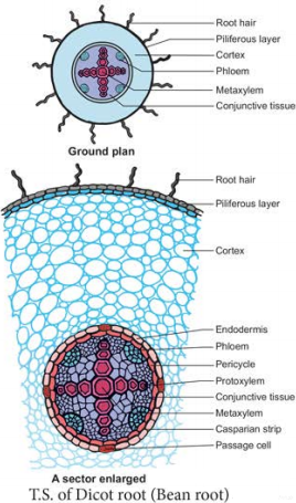
  1. Using hand cutting method to make thin slice of dicot root.
  2. To make slide and stain of plant sample.
  3. To observe the plant sample under microscope.
Materials:
  1. A young dicot root
  2. Compound microscope
  3. Slide
  4. Cover slip
  5. Eosin stain
Method:
  1. Place 2 cm of young dicot root on a glass slide or plate.
  2. Cut thin slices of the root through the region of maturation.
  3. Stain it with Eosin.
  4. Fix one or two of the sections in a slide and put a cover slip.
  5. To observe the sample under a compound microscope and record the parts of the sample.
View full question & answer
Question 25 Marks
Explain the various parts of sunflower leaf with neat diagram.
Answer
1. Anatomy of a Dicot Leaf – sunflower Leaf: Internal structure of dicotyledonous leaves reveal epidermis, mesophyll and vascular tissues.
2. Epidermis: This leaf is generally dorsiventral. It has upper and lower epidermis. The epidermis is usually made up of a single layer of cells that are closely packed. The cuticle on the upper epidermis is thicker than that of lower epidermis. The minute opening found on the epidermis are called stomata. Stomata are more in number on the lower epidermis than on the upper epidermis.
A stomata is surrounded by a pair of bean shaped cells are called guard cells. Each stoma internally opens into an air chamber. These guard cells contain chlotroplasts. The main function of epidermis is to give protection to the inner tissue called mesospyll. The cuticle helps to check transpiration. Stomata are used for transpiration and gas exchange.
3. Mesophyll: The entire tissue between the upper and lower epidermis is called mesophyll (GK meso = in the middle, phyllome = leaf). There are two regions in the mesophyll. They are palisade parenchyma and spongy parenchyma. Palisade parenchyma cells are seen beneath the upper epidermis. It consists of vertically elongated cylindrical cells in one or more layers. These are compactly arranged and are generally without intercellular spaces. Palisade parenchyma cells contain more chloroplasts than the spongy parenchyma cells. The function of palisade parenchyma is photosynthesis. Spongy parenchyma lies below the palsied parenchyma. Spongy cells are irregularly shaped. These cells are very loosly arranged with numerous airspaces. As compared to palisade cells, the spongy cells contain number of chloroplasts. Spongy cells facilitate the exchange of gases with the help of air spaces. The air space that is found next to the stomata is called respiratory cavity or substomatal cavity. Å 4. Vascular tissue: Vascular tissue are present in the veins of leaf. Vascular bundles are conjoint collateral and closed. Xylem is present towards the upper epidermis, while the phloem towards the lower epidermis. Vascular bundles are surrounded by a compact layer by a parenchymatous cells called bundle sheath or border parenchyma.Xylem consists of metaxylem and protoxylem elements. Protoxylem is present towards the upper epidermis, while the phloem consists of sieve tubes, companion cells and phloem parenchyma.
Phloem fibres are absent. Xylem sonsists of vessels and xylem parenchyma. Tracheids and xylem fibres are absent.

Image
View full question & answer
Question 45 Marks
Explain in detail about the vascular bundles of monocot stem.
Answer
1. Vascular bundles: Vascular bundles are scattered (atactostele) in the parenchyma ground tissue. Each vascular bundle is surrounded by a sheath of sclerenehymatous fibres called bundle sheath. The vascular bundles are conjoint, collateral, endarch and closed. Vascular bundles are numerous, small and closely arranged in the peripheral portion. Towards the centre, the bundles are comparatively large in size and loosely arranged. Vascular bundles are skull or oval shaped.2. Phloem: The phloem in the monocot stem consists of sieve tubes and companion cells. Phloem parenchyma and phloem fibres are absent. It can be distinguished into an outer crushed protophloem and an inner metaphloem.3. Xylem: Xylem vessels are arranged in the form of ‘Y’ the two metaxylem vessels at the base. In a mature bundle, the lowest protowylem disintegrates and forms a cavity known as protoxylem lacuna.
View full question & answer
Question 55 Marks
Draw and label the various parts of T.S. of dicot root.
Answer
Draw and label the various parts of T.S. of dicot root:
Image
View full question & answer
Question 75 Marks
Compare the different types of plant tissues.
Answer
DistributionMain functionsNatureCell shapeWall materials
ParenchymaCortex, Pith
medullary rays
and Packing
tissues in vascular
system
Packing tissue, support,
gaseous exchange,
food storage
LivingUsually IsodiametricMainly
Cellulose and Pectinase

CollenchymaOuter region
of cortex as in angles of stems, mid-rib
of leaves
MechanicalLivingElongated PolygonalMainly Cellulose, Pectin and Hemi-cellulose
Sclerenchyma

(a) Fibre
Outer region of cortex,
pericycle of
stems, vascular
bundles
MechanicalDeadElongated and
Polygonal with
tapering ends
Mainly Lignin
(b) SclereidsCortex, Pith;
Phloem shells
and stones of
fruits and seed
coats
Mechanical

Protection
DeadRoughly
Isodiametric
with much
variation
Mainly lignin
Tracheids and

Vessels
Vascular
System
Translocation
of water and
mineral salts
DeadElongated and
Tubular
Mainly lignin
Phloem Sieve

tubes
Vascular
System
Translocation
of organic
solutes
LivingElongated and

Tubular
Cellulose,
Pectir and
Hemicellulose
Companion

Cells
Vascular
System
Work in
association
with sieve
tubes
LivingElongated and
narrow
Cellulose,
Pectin and
Hemicellulose
View full question & answer
Question 85 Marks
Describe the types of tracheids with diagram.
Answer
Image
Types of secondary wall thickenings in tracheids and vessels:
Tracheids are dead, lignified and elongated cells with tapering ends. Its lumen is broader than that of fibres. In cross section, the tracheids are polygonal. There are different types of cell wall thickenings due to the deposition of secondary wall substances. They are annular (ring like), spiral (spring like), scalariform (ladder like) reticulate (net like) and pitted (uniformly thick except at pits). Tracheids are imperforated cells with bordered pits on their side walls. Only through this conduction takes place in Gymnosperms. They are arranged one above the other. Tracheids are chief water conducting elements in Gymnosperms and Pteridophytes. They also offer mechanical support to the plants.
View full question & answer
Question 95 Marks
Describe the structure and function of different kinds of parenchyma tissues?
Answer
Image
Parenchyma is generally present in all organs of the plant. It forms the ground tissue in a plant. Parenchyma is a living tissue and made up of thin walled cells. The cell wall is made up of cellulose. Parenchyma cells may be oval, polyhedral, cylindrical, irregular, elongated or armed. Parenchyma tissue normally has prominent intercellular spaces. Parenchyma may store various types of materials like, water, air, ergastic substances. It is usually colourless. The turgid parenchyma cells help in giving rigidity to the plant body. Partial conduction of water is also maintained through parenchymatous cells. Occsionaliy Parenchyma cells which store resin, tannins, crystals of calcium carbonate, calcium oxalate are called idioblasts. Parenchyma is of different types and some of them are discussed as follows. Types of paranchyma:
(i) Aerenchyma: Parenchyma which contains air in its intercellular spaces. It helps in aeration and buoyancy. eg: Nymphae and Hydrilia
Image
(ii) Storage Parenchyma: parenchyma stores food materials. eg: Root and stem tubers.
Image
(iii) Stellate Parenchyma: Star shaped parenchyma. eg: Petioles of Banana and Canna.
Image
(iv) Chlorenchyma: Parenchyma cells with chlorophyll. Function is photosynthesis, eg: Mesophyll of leaves.
Image
(v) Prosenchyma: parenchyma cells became elongated, pointed and slightly thick walled. It provides mechanical support.
Image
View full question & answer
Question 105 Marks
Explain Histogen theory, Korper Kappe Theory and Quiescent Centre Concept with diagrams.
Answer
Histogen Theory: Histogen theory is proposed by Hanstein (1868) and supported by Strassburgur. The histogen theory as appilied to the root apical meristem speaks of four histogen in the meristem. They are respectively
  1. Dermatogen: It is a outermost layer. It gives rise to root epidermis.
  2. Periblem: it is a middle layer. It gives rise to cortex.
  3. Plerome: It is innermost layer. It gives rise to stele.
  4. Calyptrogen: it gives rise to root cap.
Image
Korper Kappe Theory: Korper kappe theory is proposed by Schuepp. There are two zones in root apex – Korper and Kappe.
  1. Korper zone forms the body.
  2. Kappe zone forms The cap.
This theory is equivalent to tunica corpus theory of shoot apex.The two divisions are distinguished by the type of T (also called Y divisions). Korper is characterised by inverted T division and kappe by straight T divisions.

Image
Quiescent Centre Concept: Quiescent centre concept was proposed by Clowes (1961) to explain root apical meristem activity. These centre is located between root cap and differentiating cells of the roots. The apparently inactive region of cells in root promeristem is called quiescent centre. It is the site of hormone synthesis and also the ultimate source of all meristematic cells of the meristem.Image
View full question & answer
[5 Mark each] - BIO BOTANY STD 11 Questions - Vidyadip