Question 13 Marks
Differentiate between dendrites and axons.
Answer
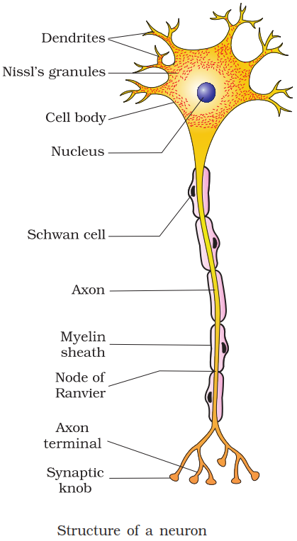
View full question & answer→Dendrites and axons:
| Dendrites | Axons | ||
| 1. | Dendrite is a small projection arising from the neuron. It conducts the nerve impulse toward the cell body. | 1. | Axon is a single, long projection that conducts the nerve impulse away from cell body to the next neuron. |
| 2. | Nissl’s granules are present in dendrites. | 2. | Nissl’s granules are absent from axons. |
| 3. | Dendrites are always non-myelinated. | 3. | Axons can be myelinated or non-myelinated. |