Question
Describe the respiratory system of human.

Answer

Respiratory system of human : Human respiratory system consists of nostrils, nasal chambers, pharynx, larynx, trachea, bronchi, bronchioles, lungs, diaphragm and intercostal muscles.
1. Nostrils and nasal chambers:
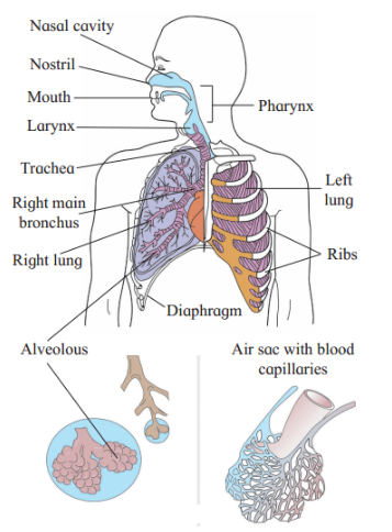
Image

  1. Oxygen rich air is taken in the body through the nostrils or external nares. They are external opening of the nose. Carbon dioxide and water vapour are also released out of the body through the same passage i.e. the nostrils.
  2. Internal nares open into the pharynx. The space between external and internal nares is knows as nasal chamber which is lined internally by mucous membrane and ciliated epithelium.
  3. Nasal chamber is divided into two parts by a cartilage called mesethmoid. Each part of these halves is further divided into three regions, viz. vestibule, respiratory part and sensory part.
  4. Vestibule is the anteriormost part of nasal chamber. In the vestibule fine hairs are present. They filter out the dust particles and prevent them from going inside.
  5. Respiratory part is the second region which is richly supplied with the capillaries. Air is made warm and moist in this region.
  6. Sensory part is lined by sensory epithelium. It is concerned with the detection of smell.

2. Pharynx:

  1. Pharynx is a short and vertical tube measuring about 12 cm in length. In pharynx the respiratory and food passages cross each other.
  2. The upper part of pharynx is known as naso-pharynx which conducts the air. The lower part is called laryngo-pharynx or oro¬pharynx which conducts food to the oesophagus.
  3. Tonsils that are made up of lymphatic tissue are present in the pharynx. They kill the bacteria by trapping them in the mucus.

3. Larynx:

  1. Larynx produces sound. In males it increases in size at puberty. This is termed as Adam’s apple. It is clearly seen in the neck region.
  2. From pharynx air enters the larynx. The opening through which it enters is called glottis. Glottis is guarded by a flap called epiglottis.
  3. Epiglottis prevents the entry of food particles into the trachea.
  4. TWo folds of elastic tissue called vocal cords are seen along the side of glottis. When they vibrate the sound is produced.

4. Trachea:

  1. The trachea or wind pipe is about 12 cm long and 2.5 cm wide.
  2. It is situated in front of the oesophagus and runs downwards in the thorax through the neck.
  3. The trachea is made up of fibrous muscular tissue wall which is supported by ‘C’-shaped cartilages. These cartilaginous rings are 16 to 20 in number.
  4. Internally the tracheal wall bears ciliated epithelium and mucous glands.
  5. When any foreign particle enters the trachea inadvertently. It is thrown out by coughing action.
  6. Mucous and ciliary action remove the dust particles and push them upwards to the larynx. These particles are then gulped and taken into the oesophagus.

5. Bronchi and bronchioles:

  1. At the distal end, the trachea divides into two bronchi (Singular – bronchus). Bronchi lie below the sternum or breast bone.
  2. Each bronchus has a complete ring of cartilage for support. The two bronchi enter into the lungs on either side.
  3. After entering into the lungs each bronchus divides into secondary and tertiary bronchi. The tertiary bronchi divide and re-divide to form minute bronchioles.
  4. Bronchioles do not have cartilages in their walls. Each bronchiole ends into a balloon like alveolus.
  5. Owing to the presence of alveoli the lungs become spongy and elastic.

6. Lungs:

  1. Lungs are principal respiratory organs located in the thoracic cavity.
  2. They are pinkish, soft, hollow, paired, elastic and distensible organs.
  3. Each lung is enclosed in a pleural sac which consists of two membranes, viz. an outer parietal and inner visceral.
  4. The parietal and visceral membranes enclose pleural cavity which is filled with pleural fluid. The pleural fluid lubricates and prevents friction when pleural membranes slide on each other.
  5. Lungs are highly vascular as they are richly supplied with blood capillaries.
  6. The left lung has two lobes while the right lung has three lobes. Each lobe has many bronchioles and alveolar sacs. The alveolar sacs are spherical and thin walled.
  7. Each alveolar sac contains about 20 alveoli. The alveoli appear as a bunch of grapes. The lobule in the lung thus consists of alveolar ducts, alveolar sacs and alveoli.
  8. Each alveolus has thin and elastic walls. It is about 0.1 mm in diameter. Alveoli are covered by network of capillaries from pulmonary artery and pulmonary vein. A network of pulmonary capillaries supply the alveolus.
  9. The alveolar wall is 0.0001 mm thick and made up of simple, non-ciliated, squamous epithelium. It has collagen and elastin fibres.
  10. Every lung has about 700 million alveoli. They increase the surface area of the lungs for exchange of gases.


Need a full question paper?

Generate a complete, print-ready paper with questions like this in minutes — across 16+ boards, with answer keys.

Start Generating Free

Similar questions

Discuss the various types of positive interactions between species.
Describe in detail the external genitalia of human female reproductive system.
Mortality.
Allopatric and Sympatric speciation.
Answer the following questions:
State and explain the law of dominance as proposed by Mendel.
Enlist human endocrine glands.
Describe the T.S. of thyroid gland and add a note on deficiency of thyroxine
Name the improved characteristics of wheat that helped India to achieve green revolution.
When does oogenesis start in humans? Name the three hormones and their source, linked with oogenesis. How do they influence the process of oogenesis? Explain.
Read the following and answer any four questions from (i) to (v) given below:
Crown gall is a neoplastic disease of most dicotyledonous plants and is caused by the soil bacterium Agrobacterium tumefaciens. A large extra chromosomal plasmid in these bacteria was found to be responsible for this disease. The plasmid is known as Ti plasmid. Bacteria free crown gall cells can be cultured in the absence of phytohormones. Ti plasmid is widely used in genetic engineering to deliver the desirable genes. The part of Ti plasmid transferred into plant cell DNA is called T-DNA. T-DNA with desired DNA segment is inserted into the chromosome of the host plant where it produces copies of itself.
  1. Which of the following is the full full of T-DNA?
  1. Transfer DNA.
  2. Tumour inducing DNA.
  3. Transgenic DNA.
  4. None of these.
  1. Ti plasmid cannot infect and develop crown gall in,
  1. Tomato.
  2. Maize.
  3. Soybean.
  4. sunflower.
  1. While making transgenic plant, T-DNA is inserted into the host cell, it is then:
  1. Integrated into the target host genome.
  2. Lie independent of the host genome.
  3. Rupture the host cell wall.
  4. Produce oncogenic factors in the host.
  1. Agrobacterium mediated gene transfer in plants:
  1. Allows relatively large segment of DNA.
  2. Transfer of DNA with defined ends and minimal rearrangement.
  3. High quality and fertility in transgenic plants.
  4. All of these.
  1. Assertion: In Agrobacterium mediated gene transfer in plants, transgenic plants do not develop tumors.
Reason: In T-DNA tumor producing genes are deleted during the process of gene transfer.
  1. Both assertion and reason are true and reason is the correct explanation of assertion.
  2. Both assertion and reason are true but reason is not the correct explanation of assertion.
  3. Assertion is true but reason is false.
  4. Both assertion and reason are false.
A large number of married couples the world over are childless. It is shocking to know that in India the female partner is often blamed for the couple being childless.
  1. Why in your opinion the female partner is often blamed for such situations in India? Mention any two values that you as a biology student can promote to check this social evil.
  2. State any two reasons responsible for the cause of infertility.
  3. Suggest a technique that can help the couple to have a child where the problem is with the male partner.