Heterotypic division is first meiotic division, during which a diploid cell is divided into two haploid cells. The daughter cells resulting from this division are different from the parent cell in chromosome number. Hence the division is called heterotypic division.It consists of following phases:1. Prophase -I:It is the most complicated and longest phase of meiotic division.It is further divided into five sub-phases viz. leptotene, zygotene, pachytene, diplotene and diakinesis.a. Leptotene:
b. Zygotene:
c. Pachytene:
d. Diplotene:The chiasma becomes clearly visible in diplotene due to beginning of repulsion between synapsed homologous chromosomes. This is known as desynapsis. Synaptonemal complex also starts to disappear. e. Diakinesis:
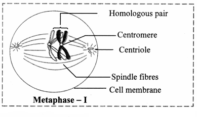
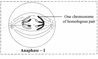
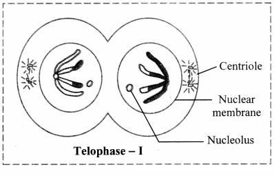
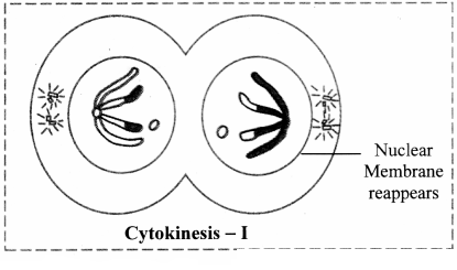
2. Metaphase -1:a. The spindle fibres are well developed.b. The tetrads orient themselves on equator in such a way that centromeres of homologous tetrads lie towards the poles and arms towards the equator.c. They are ready to separate as repulsive force increases.a. Homologous chromosomes are carried towards the opposite poles by spindle apparatus. This is known as disjunction.b. The two sister chromatids of each chromosome do not separate in meiosis -I. This is reductional division.c. The sister chromatids of each chromosome are connected by a common centromere.d. Both sister chromatids of each chromosome are now different in genetic content as one of them has undergone recombination.3. Anaphase – I:1. Homologous chromosomes are carried towards the opposite poles by spindle apparatus. This is known as disjunction.2. The two sister chromatids of each chromosome do not separate in meiosis -I. This is reductional division.3. The sister chromatids of each chromosome are connected by a common centromere.4. Both sister chromatids of each chromosome are now different in genetic content as one of them has undergone recombination.4. Telophase-I:a. The haploid number of chromosomes becomes uncoiled and elongated after reaching their respective poles.b. The nuclear membrane and nucleolus reappear and thus two daughter nuclei are formed.Cytokinesis -1:Cytokinesis occurs after karyokinesis and two haploid cells are formed. In many cases, these daughter cells pass through interkinesis.[Note: The association between the homologous chromosomes i.e. chiasmata remain till metaphase I. During metaphase /, the paired homologous chromosomes move to the metaphase plate. In anaphase [ the spindle fibers begin to shorten. As these spindle fibres shorten, the association between homologous chromosomes (chiasmata) are broken, allowing homologous chromosomes to be pulled to opposite poles.]
Generate a complete, print-ready paper with questions like this in minutes — across 16+ boards, with answer keys.