Question 15 Marks
Write down the explanation of prophase I in your own words.
Prophase -I:
Leptotene:
Zygotene:
Pachytene:
Diplotene:
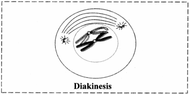
Diakinesis:
Prophase -I:
Leptotene:
Zygotene:
Pachytene:
Diplotene:
Diakinesis:
Answer
View full question & answer→ANS 1 It is the most complicated and longest phas0e of meiotic division.
It is further divided into five sub-phases viz. leptotene, zygotene, pachytene, diplotene and diakinesis.
ANS 2
ANS 6
It is further divided into five sub-phases viz. leptotene, zygotene, pachytene, diplotene and diakinesis.
ANS 2
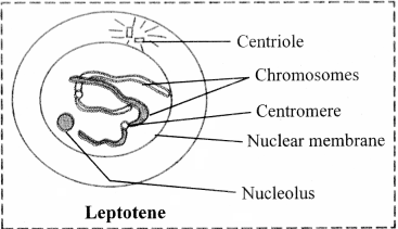
- The volume of the nucleus increases.
- The chromosomes become long distinct and coiled.
- They orient themselves in a specific fonn known as bouquet stage. This is characterized with the ends of chromosomes converged towards the side of nucleus where the centrosome lies.
- The centriole duplicates into two and migrates to opposite poles. [Note: Centrioles divide during $G_j$ phase of interphase.]
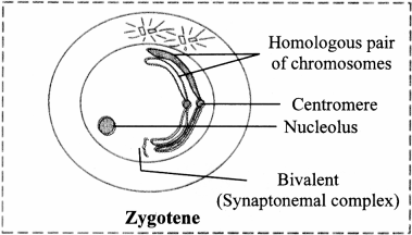
- Pairing of non-sister chromatids of homologous chromosomes takes place by formation of synaptonemal complex. This pairing is called synapsis.
- Each pair consists of a maternal chromosome and a paternal chromosome. Chromosomal pairs are called bivalents or tetrads.
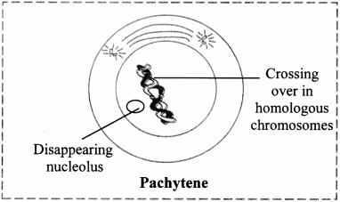
- Each individual chromosome begins to split longitudinally into two similar chromatids. Therefore, each bivalent now appears as a tetrad consisting of four chromatids.
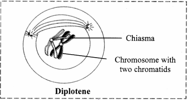
- The homologous chromosomes begin to separate but they do not separate completely and remain attached to one or more points.
- These points are called chiasmata (Appear like a cross-X).
- Chromatids break at these points and broken segments are exchanged between non-sister chromatids of homologous chromosomes resulting in recombination.
ANS 6
- The chiasmata begin to move along the length of chromosomes from the centromere towards the ends of chromosomes. The displacement of chiasmata is termed as terminalization.
- The terminal chiasmata exist till the metaphase.
- The nucleolus and nuclear membrane completely disappear and spindle fibres begin to appear.