Question
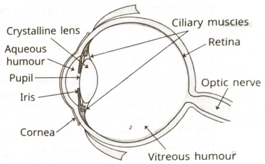
Draw cross-sectional lebelled diagram of human eye. Describe its construction and function of each part. Also describe how we can see through our eyes.

Answer

Image
→ Human eye is like a camera. Its lens system forms a real and inverted image on the rear light sensitive screen, called "retina." $\Rightarrow$ → Light enters the eye through a thin transparent membrane called the "cornea." If forms the transparent bulge on the front surface of an eye ball as shown in above figure.
→ Eyeball is nearly spherical in shape with diameter approximately $2.3 cm$. Most of the refraction of light rays entering the eye takes place at the outer surface of comea. The crystalline lens merely provides fine adjustment of focal length which is required to produce images of objects located at different distances, on the retina.
→ Just behind the cornea, there is a dark muscular diaphragm, called "iris" which controls the size of "pupil" (an opening in the center of iris which gives colour to our eyes.)
→ The pupil regulates and controls the amount of light which enters in our eye.
→ The eye lens forms an inverted real image of every object on the retina (inner back surface of eye). The retina is a delicate membrane having enormous no. of light sensitive cells, which get activated upon the incidence of light and then they generate electrical signals. These signals are immediately sent to brain via optic nerves. The brain interprets (understands) these signals, processes the information and finally we see the objects as they are.
→ There are two types of cells in the retina
(i) Rods : These cells are sensitive to low intensity light.
(ii) Cones : These cells are sensitive to high intensity light. They help us to identify different colours.

Need a full question paper?

Generate a complete, print-ready paper with questions like this in minutes — across 16+ boards, with answer keys.

Start Generating Free