Questions

Case study (4 Marks)

Take a timed test

7 questions · self-marked practice — reveal the answer and mark yourself.

Question 14 Marks
Explain the amplification of gene of interest using PCR (Diagram is must)
View full question & answer
Question 24 Marks
Explain pBR322 with its cloning sites, enzymes with well labeled diagram.###Explain: cloning sites. (Diagram is not necessary)
Answer
Cloning sites :
→ In order to link the alien DNA, the vector needs to have very few, preferably single, recognition sites for the commonly used restriction enzymes.
→ Presence of more than one recognition sites with in the vector will generate several fragments, which will complicate the gene cloning.
Image
→ The ligation of alien DNA is carried out at a restriction site present in one of the two antibiotic resistance genes.
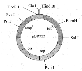
→ For example, you can ligate a foreign DNA at the BamHI site of tetracycline resistance gene in the vector pBR322.
→ The recombinant plasmids will lose tetracycline resistance due to insertion of foreign DNA but can still be selected out from non-recombinant ones by plating the transformants on tetracycline containing medium.
→ The transformants growing on ampicillin containing medium are then transferred on a medium containing tetracycline.
→ The recombinants will grow in ampicillin containing medium but not on that containing tetracycline.
→ But, non recombinants will grow on the medium containing both the antibiotics.
→In this case, one antibiotic resistance gene helps in selecting the transformants, whereas the other antibiotic resistance gene gets 'inactivated due to insertion' of alien DNA, and helps in selection of recombinants.
View full question & answer
Question 34 Marks
Explain: separation and isolation of DNA fragments.###How will you make separation and isolation of DNA fragments by using gel electrophoresis?
Answer
→ The techniques used for separation and isolation of DNA fragments is known as gel electrophoresis.
→ In this method Agarose gel is used as a medium.
→ The cutting of DNA by restriction endonucleases results in the fragments of DNA.
→ These fragments can be separated by a technique as shown in figure.
Image
→ Since, DNA fragments are negatively charged molecules they can be separated by forcing them to move towards the anode under an electric field througha medium/matrix.
→ The DNA fragments separate (resolve) according to their size through sieving effect provided by the agarose gel.
→ Hence, the smaller the fragment size, the farther it moves.
→ The separated DNA fragments can be visualised only after staining the DNA with a compound known as ethidium bromide followed by exposure to UV radiation.
→ DNA appears as bright orange coloured bands of DNA in an ethidium bromide stained gel exposed to UV light.
→ The separated bands of DNA are cut out from the agarose gel and extracted from the gel piece. This step is known as elution.
→ The DNA fragments purified in this way are used in constructing recombinant DNA by joining them with cloning vectors.
View full question & answer
Question 44 Marks
Explain- Restriction enzymes.
Answer
Empty → In the year 1963, the two enzymes responsible for restricting the growth of bacteriophage in Escherichia coli were isolated.
→ One of these added methyl groups to DNA, while the other cut DNA.
→ The later was called restriction endonuclease.
→ The first restriction endonuclease Hind - II, whose functioning depended on a specific DNA nucleotide sequence.
→ Hind -II was isolated and characterised five years later.
→ It was found that Hind II always cut DNA molecules at a particular point by recognising a specific sequence of six base pairs.
→ This specific base sequence is known as the recognition sequence for Hind II.
→ Besides Hind II, today we know more than 900 restriction enzymes that have been isolated from over 230 strains of bacteria each of which recognise different recognition sequences.
Nomenclature :
→ The convention for naming these enzymes is the first letter of the name comes from the genus and the second two letters come from the species of the prokaryotic cell from which they were isolated.
e.g., EcoRI
→ It comes from Escherichia coli RY 13.
→ In EcoRI, the letter 'R' is derived from the name of strain.
→ Roman numbers following the names indicate the order in which the enzymes were isolated from that strain of bacteria.
TYPES :
→ Restriction enzymes belong to a larger class of enzymes called nucleases. These are of two kinds.
(a) Exonucleases
(b) Endonucleases.
(1) Exonucleases remove nucleotides from the ends of the DNA.
(2) Endonucleases make cuts at specific positions within the DNA.
→ Each restriction endonuclease functions by 'inspecting' the length of a DNA sequence.
→ Once it finds its specific recognition sequence, it will bind to the DNA and cut each of the two strands of the double helix at specific points in their sugar -phosphate backbones.
PALINDROMIC SEQUENCE :
→ Each restriction endonuclease recognises a specific palindromic nucleotide sequences in the DNA.
→ The palindrome in DNA is a sequence of base pairs that reads same on the two strands when orientation of reading is kept the same. For example, the following sequences reads the same on the two strands in 5'→3' This is also true if read in the 3' → 5' direction. 5' direction.
5' ____ GAATTC _____ 3'
3' ____ CTTAAG _____ 5'
Mechanism of action :
→ Restriction enzymes cut the strand of DNA a little away from the centre of the palindrome sites, but between the same two bases on the opposite strands.
→ This leaves single stranded portions at the ends.
→ There are overhanging stretches called sticky ends on each strand.
Image
→ When cut by the same restriction enzyme, the resultant DNA fragments have the same kind of 'sticky-ends' and, these can be joined together using DNA ligases.
→ Unless one cuts the vector and the source DNA with the same restriction enzyme, the recombinant vector molecule cannot be created.
→ Restriction endonucleases are used in genetic engineering to form 'recombinant' molecules of DNA, which are composed of DNA from different sources/genomes.
View full question & answer
Question 54 Marks
How interesingly Gene is amplified by using PCR? Explain with diagram.
Answer
→ PCR stands for Polymerase Chain Reaction.
→ In this reaction, multiple copies of the gene (or DNA) of interest is synthesised in vitro using two sets of primers (small chemically synthesised oligonucleotides that are complementary to the regions of DNA) and the enzyme DNA polymerase.
→ The enzyme extends the primers using the nucleotides provided in the reaction and the genomic DNA as template.
→ If the process of replication of DNA is repeated many times, the segment of DNA can be amplified to approximately billion times, i.e., 1 billion copies are made.
→ Such repeated amplification is achieved by the use of a thermostable DNA polymerase (isolated from a bacterium, Thermus aquaticus), which remain active during the high temperature induced denaturation of double stranded DNA.
→ The amplified fragment if desired can now be used to ligate with a vector for further cloning.
Image
View full question & answer
Question 64 Marks
Explain bioreactor with diagram.
View full question & answer
Question 74 Marks
Explain the methods for making host cell competent to insert the r DNA.
Answer
→ Since DNA is a hydrophilic molecule, it cannot pass through cell membranes.
→ In order to force bacteria to take up the plasmid, the bacterial cells must first be made 'competent' to take up DNA.
Micro-injection :
→ In a method known as micro-injection, recombinant DNA is directly injected into the nucleus of an animal cell.
Biolistics/ Particle bombardment :
→ In another method, suitable for plants, cells are bombarded with high velocity micro- particles of gold or tungsten coated with DNA in a method known as biolistics or gene gun.
Conformational change in the cell wall :
→ This is done by treating them with a specific concentration of a divalent cation, such as calcium, which increases the efficiency with which DNA the bacterium through pores in its cell wall.
→ Recombinant DNA can then be forced into such cells by incubating the cells with recombinant DNA on ice, followed by placing them briefly at 42°C (heat shock), and then putting them back on ice.
→ This enables the bacteria to take up the recombinant DNA.

View full question & answer