Questions

[5 Mark Question Answer]

🎯

Test yourself on this topic

6 questions · timed · auto-graded

Question 15 Marks
Differentiate between:
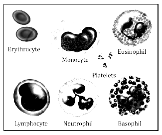
Red blood cells and White blood cells
Answer
Red blood cells White blood cells.
These are minute biconcave disc like structures, flat in centre, thick and round at the periphery and nuclei are absent in mature RBCs. These collect blood from different organs of the body.
These contain a respiratory pigment haemoglobin. Haemoglobin is not present.
RBCs help in the transport of oxygen and CO2. WBCs help in the protection of the body against infections from the germs.
The number of RBCs in an adult male is 5 million per cu. millimeter. Their number is usually about 4000-8000 per cubic millimeter.
Their average life span is about 120 days. Their average life span is about two weeks.
View full question & answer
Question 25 Marks
The figure below represents the internal structure of a mammalian heart and the associated blood vessels.

(i) (a) Name each of the structures labeled 1, 2, 3, 4, 5, 6, 7 and 8.
(b) State the function of each of the structures 5, 6, 7, and 8.
(ii) (a) State the function of the heart as an entire organ.
(b) Why are the walls of the left ventricle more muscular than the right?
Answer
(i) (a)
1. Right auricle,
2. Left ventricle,
3. Pulmonary artery,
4. Pulmonary vein,
5. Inferior vena cava,
6. Aorta,
7. Pulmonary semilunar valve,
8. Tricuspid valve.
(b)
5 — carry deoxygenated blood from the body parts to the right auricle of the heart.
6 — distributes blood all over the body.
7 — prevents the backflow of the blood into the ventricles.
8 — prevents the reverse flow of the blood from the right ventricle into the right auricle,
(ii) (a) The heart makes the blood to circulate all over the body.
(b) The walls of the left ventricle are more muscular because they have to pump blood to a larger distance than the right so that walls have to withstand high pressure.
View full question & answer
Question 35 Marks
The diagram given below represents the human heart in one phase of its activity. Study the same and then answer the questions that follow:

(i) Name the phase.
(ii) Which parts of the heart are contracting in this phase ? Give a reason to support your answer.
(iii) Name the part numbered 1 to 6.
(iv) What type of blood flows through the parts marked ‘1’ and ‘2’?
(v) How many valves are closed in this phase?
Answer
(i) Ventricular systole.
(ii) Both ventricles are contracting in this phase, because both bicuspid and tricuspid valves are closed in order to prevent the back flow of blood into auricles and the semilunar valves are open.
(iii) 1. Pulmonary artery,
2. Aorta,
3. Pulmonary vein,
4. Left auricle,
5. Bicuspid valve (mitral valve),
6. Right ventricle.
(iv)
‘1’—carries deoxygenated blood.
‘2’—carries oxygenated blood.
(v) Two (Bicuspid and Tricuspid) valves.
View full question & answer
Question 45 Marks
Given below is a simple diagram of the circulation of blood in a mammal showing the main blood vessels, the heart, lungs, and body tissues. The blood vessel labeled 6 contains deoxygenated blood and 2 the valve leading to it has three semi-lunar pockets.

(i) Name the blood vessels of organs marked by numbers 1 to 8.
(ii) What do you mean by the term ‘double circulation’ of blood in mammals?
(iii) What is diastole?
Answer
(i)
1. Veins from tissues.
2. Superior vena cava
3. Aorta
4. Right ventricle
5. Left ventricle
6. Pulmonary Artery
7. Pulmonary vein
8. Lungs.
(ii) The heart is said to have double circulation because the blood passes through the heart twice.
(a) It first leaves t.rough the right ventricle, goes to the lungs and then returns to the left auricle of the heart (pulmonary circulation).
(b) It leaves through the left ventricle, circulates through the body, and again returns to the right auricle of heart (systemic circulation).
(iii) Diastole is the phase of relaxation of the heart muscles during which the heart chambers fill with blood and the supply of blood to the cardiac muscle is improved.
View full question & answer
Question 55 Marks
The diagram below represents the simplified pathway of the circulation of blood. Study the same and answer the questions which follow:

(i) Name the blood vessels labelled 1 and 2.
(ii) State the function of blood vessels labelled 5 and 8.
(iii) What is the importance of the blood vessel labelled 6?
(iv) Which blood vessel will contain a high amount of glucose and amino acids after a meal?
(v) Draw a diagram of the different blood cells as seen in a smear of human blood.
Answer
(i)
1- Superior vena cava
2 - Aorta
(ii) Function of blood vessel 5 (Hepatic artery) - It supplies oxygenated blood to the liver.
Function of blood vessel 8 (Inferior vena cava) - It carries deoxygenated blood from the posterior parts of the body to the heart.
(iii) Importance of blood vessel 6 (Hepatic portal vein) - It carries nutrient laden blood from the gastrointestinal tract to the liver.
(iv) Blood vessel 6, i.e. hepatic portal vein.
(v) Different blood cells seen in a smear of human blood:
View full question & answer
Question 65 Marks
The diagram represents the ‘closed system’ or ‘double circulation’ of blood in mammals.

Justify the above statement.
State two structural and two functional differences between the arteries and veins.
State the changes in the composition of blood as it passes through the following organs:
(a) Lungs (b) Gut (c) Liver (d) Kidneys.
Answer
Double Circulation or closed system:
In human beings, the heart has double circulation as the blood passes through the heart twice.
(1) It first leaves the heart (right ventricle) and goes to the lungs and then returns to the heart (left auricle).
(2) From the left auricle, it circulates through the entire body and again returns to the heart (right auricle).
See Answer to Question 11, differentiate between.
(a) In the lungs, the blood releases carbon dioxide and picks up oxygen.
Hence before passing through the lungs, it contains more $CO_2$​​​​​​​ while it contains more oxygen after it has passed through the lungs.
(b) When blood passes through the gut, it absorbs digested food materials i.e., it contains more carbohydrates, proteins, fats, water, ions, and vitamins.
(c) As the blood passes through the liver, it contains more urea another waste materials.
(d) As the blood passes through kidneys, it contains less urea and other waste materials.
View full question & answer
[5 Mark Question Answer] - Biology STD 10 Questions - Vidyadip