Question 15 Marks
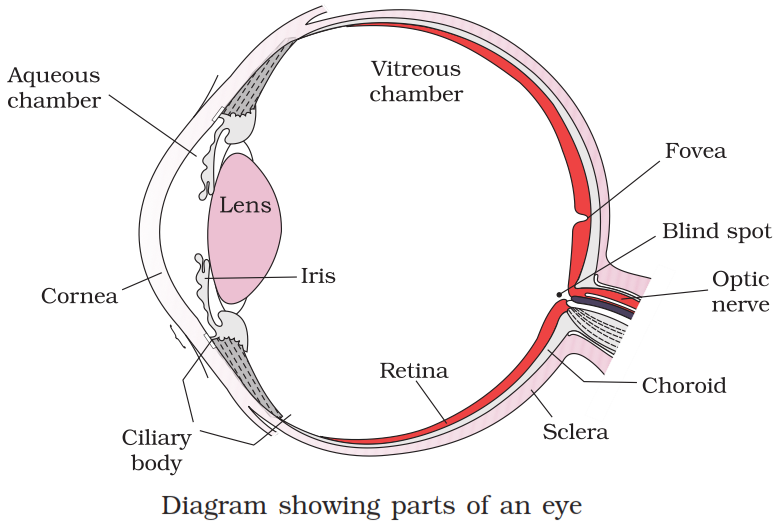
Draw labelled diagram of Eye.
35 questions · self-marked practice — reveal the answer and mark yourself.
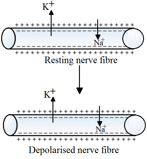

| Myelinated axons | Non-myelinated axons | ||
| 1. | Transmission of nerve impulse is faster. | 1. | Transmission of nerve impulse is slower. |
| 2. | Myelinated axon has a myelin sheath. | 2. | Myelin sheath is absent. |
| 3. | Node of Ranvier is present between adjacent myelin sheaths. | 3. | Node of Ranvier is absent. |
| 4. | Found in the brain, the spinal cord, the cranial and spinal nerves. | 4. | Found in autonomous and somatic neural systems. |
| 5. | Schwann cells are observed inside the myelin sheath. | 5. | Schwann cells are not observed inside the myelin sheath. |
|
A.
|
Resting potential
|
i.
|
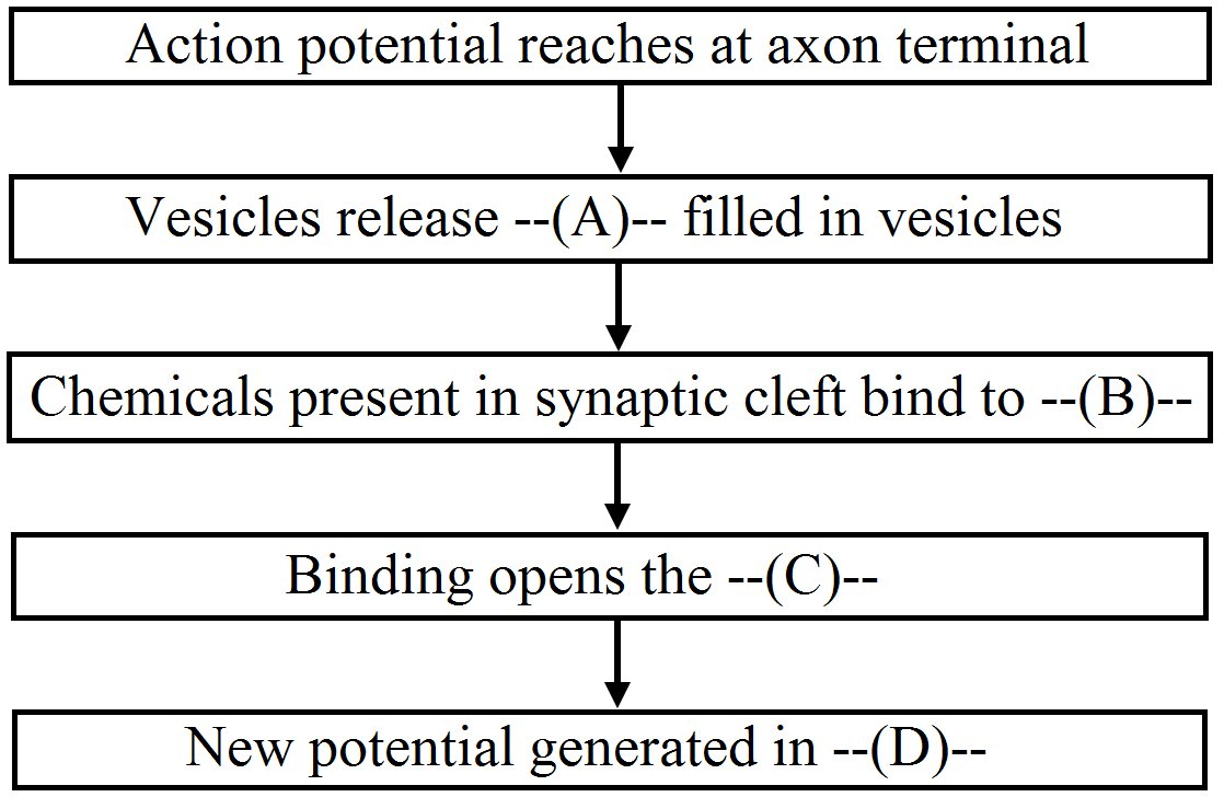
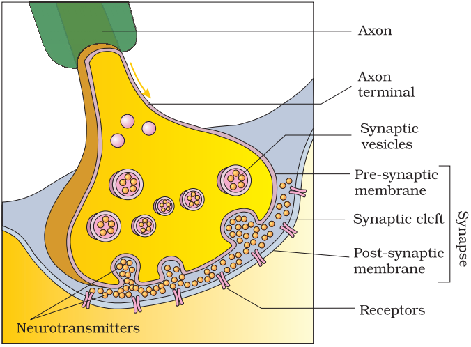
Chemicals involved in the transmission of impulses at synapses.
|
|
B.
|
Nerve impulse
|
ii.
|
Gap between the pre synaptic and post synaptic neurons.
|
|
C.
|
Synaptic cleft
|
iii.
|
Electrical potential difference across the resting neural membrane.
|
|
D.
|
Neurotransmitters
|
iv.
|
An electrical wave like response of a neuron to a stimulation.
|
|
A.
|
Resting potential
|
iii.
|
Electrical potential difference across the resting neural membrane.
|
|
B.
|
Nerve impulse
|
iv.
|
An electrical wave like response of a neuron to a stimulation.
|
|
C.
|
Synaptic cleft
|
ii.
|
Gap between the pre synaptic and post synaptic neurons.
|
|
D.
|
Neurotransmitters
|
i.
|
Chemicals involved in the transmission of impulses at synapses.
|
|
S. No.
|
Electrical Synapses
|
Chemical Synapses
|
|
1.
|
The membranes of the pre-synaptic and post synaptic neurons are in close proximity and there is no synaptic cleft.
|
The membranes of the pre-synaptic and post-synaptic neurons are separated by a fluid-filled space, the synaptic cleft.
|
|
2.
|
Electrical current can flow directly from one neuron to the other.
|
Transmission involves chemicals, called neurotransmitters.
|
|
3.
|
Impulse conduction is faster.
|
Impulse conduction is relatively slower.
|
|
4.
|
Electrical synapses are rare in our system.
|
Chemical synapses are the most common type of synapses.
|
|
|
Electrical transmission
|
Chemical transmission
|
|
i.
|
Electrical synapse is present.
|
Chemical synapase is present.
|
|
ii.
|
Membranes of pre-and post-synaptic neurons are in very close proximity provided by the gap junction.
|
The membranes of pre-and post-synaptic neurons are separated by a fluid-filled space called synaptic cleft.
|
|
iii.
|
Impulse transmission across an electrical synapse is fast.
|
Impulse transmission across a chemical synapse is slow.
|
|
iv.
|
No neurptransmitter is involved.
|
Neurotransmitter is involved.
|
|
v.
|
Electrical synapase are relatively rare.
|
Chemical synapses are relatively more.
|
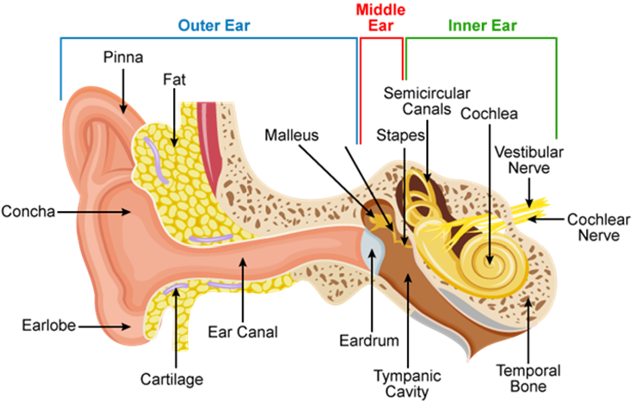
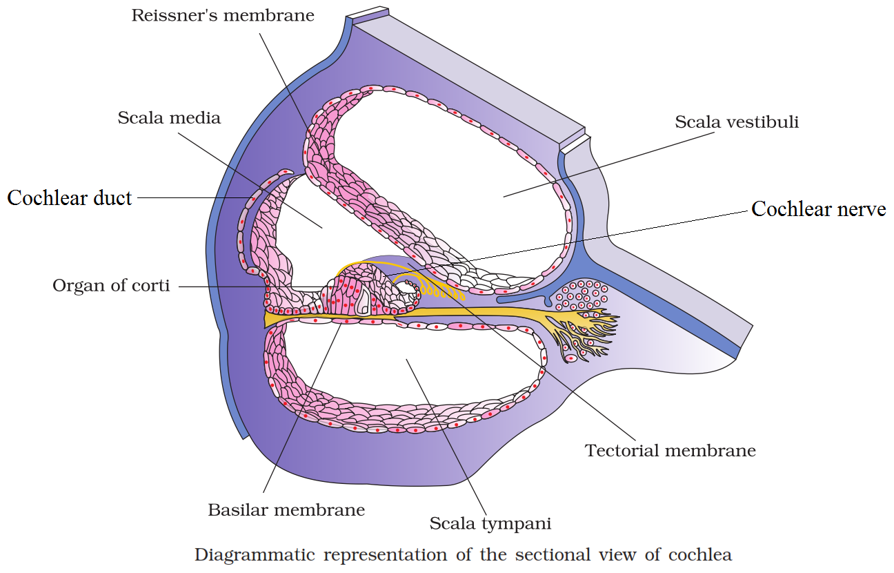
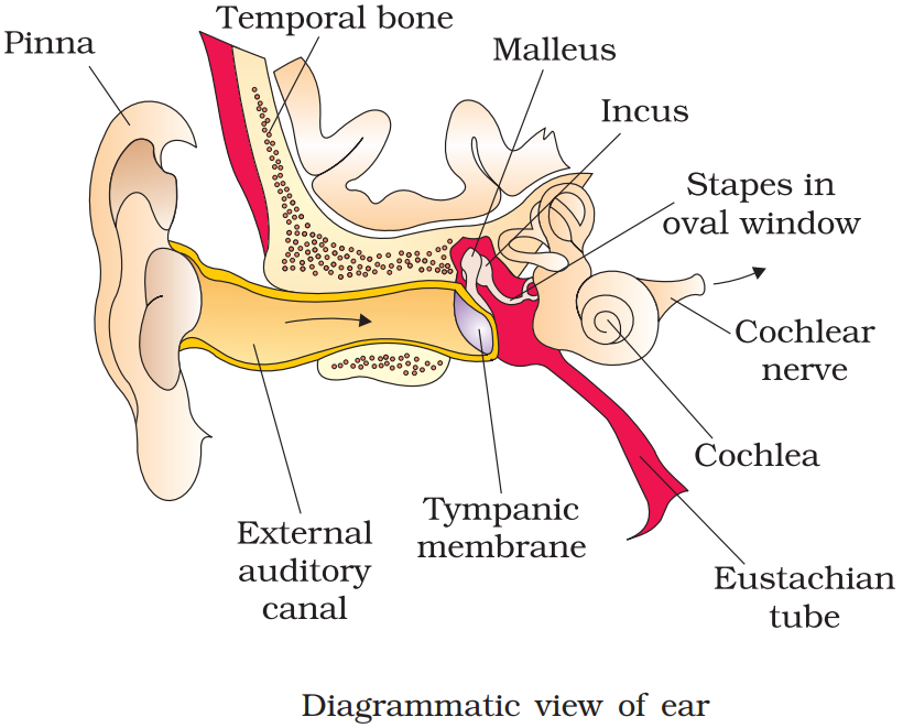
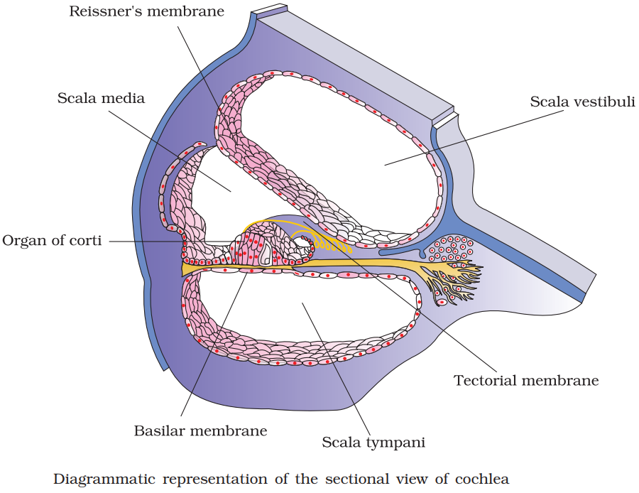
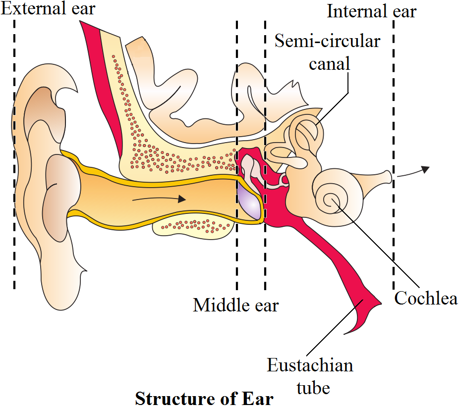 Inner Ear: The fluid-filled inner ear called labyrinth consists of two parts, the bony and the membranous labyrinths. The bony labyrinth is a series of channels Inside these channels lies the membranous labyrinth, which is surrounded by a fluid called perilymph. The membranous labyrinth is filled with a fluid called endolymph. The coiled portion of the labyrinth is called cochlea. The membranes constituting cochlea, the issner's and basilar, divide the surounding perilymph filled bony labyrinth into an upper scala vestibuli and a lower scala tympani. The space within cochlea called scala media is filled with endolymph. At the base of the cochlea, the scala vestibuli ends at the oval window, while the scala tympani terminates at the round window which opens to the middle ear. The organ of corti is a structure located on the basilar membrane which contains hair cells that act as auditory receptors. The hair cells are present in rows on the internal side of the organ of corti. The basal end of the hair cell is in close contact with the afferent nerve fibres. A large number of processes called stereo cilia are projected from the apical part of each hair cell. Above the rows of the hair cells is a thin elastic membrane called tectorial membrane. The inner car also contains a complex system called vestibular apparatus, located above the cochlea. The vestibular apparatus is composed of three semi-circular canals and the otolith organ consisting of the saccule and utricle. Each semicircular canal lies in a different plane at right angles to each other. The membranous canals are suspended in the perilymph of the bony canals. The base of canals is swollen and is called ampulla, which contains a projecting ridge called crista ampullaris which has hair cells. The saccule and utricle contain a projecting ridge called macula. The crista and macula are the specifice receptors of the vestibular apparatus responsible for maintenance of balance of the body and posture.
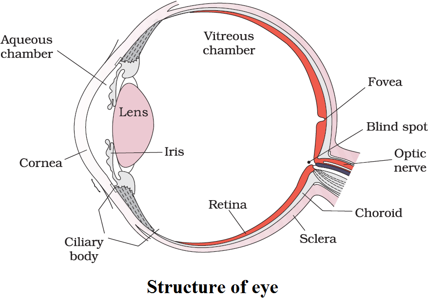 The inner layer is the retina and it contains three layers of cells from inside to outside - ganglion cells, bipolar cells and photoreceptor cells. There are two types of photoreceptor cells, namely, rods and cones. These cells contain the light-sensitive proteins called the photopigments. The daylight (photopic) vision and colour vision are functions of cones and the twilight (scotopic) vision is the function of the rods. The rods contain a purplish-red protein called the rhodopsin or visual purple, which contains a derivative of Vitamin A. In the human eye, there are three types of cones which possess their own characteristic photopigments that respond to red, green and blue lights. The sensations of different colours are produced by various combinations of these cones and their photopigments. When these cones are stimulated equally, a sensation of white light is produced. The optic nerves leave the eye and the retinal blood vessels enter it at a point medial to and slightly above the posterior pole of the eye ball. Photoreceptor cells are not present in that region and hence it is called the blind spot. At the posterior pole of the eye lateral to the blind spot, here is a yellowish pigmented spot called macula lutea with a central pit called the fovea. The fovea is a thinned-out portion of the retina where only the cones are densely packed. It is the point where the visual acuity resolution) is the greatest. The space between the cornea and the lens is called the aqueous chamber and contains a thin watery fluid called aqueous humor. The pace between the lens and the retina is called the vitreous chamber and is filled with a transparent gel called vitreous humor.