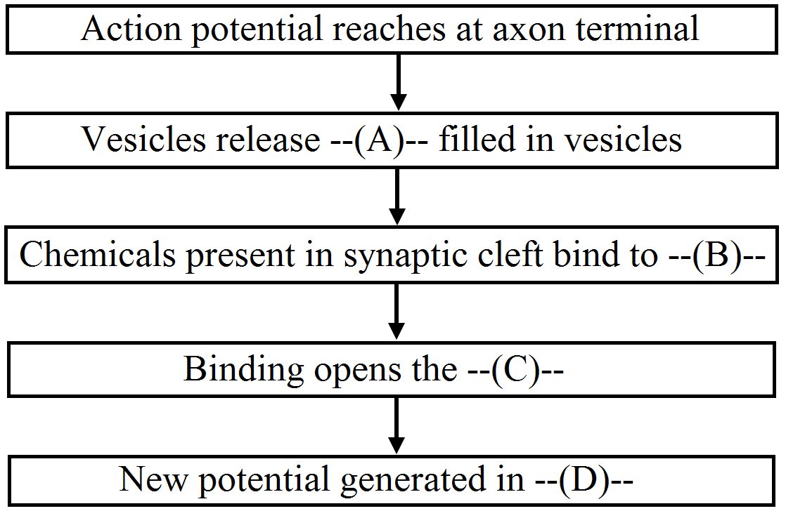
Question
Describe the detail structure of the human ear.
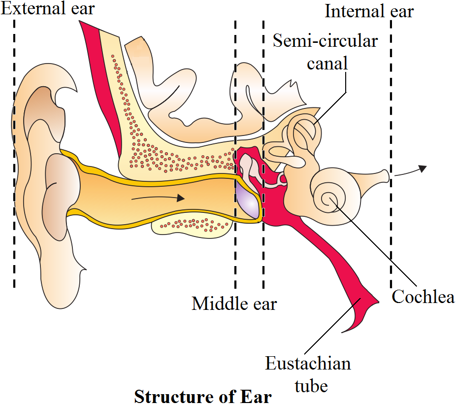 Inner Ear: The fluid-filled inner ear called labyrinth consists of two parts, the bony and the membranous labyrinths. The bony labyrinth is a series of channels Inside these channels lies the membranous labyrinth, which is surrounded by a fluid called perilymph. The membranous labyrinth is filled with a fluid called endolymph. The coiled portion of the labyrinth is called cochlea. The membranes constituting cochlea, the issner's and basilar, divide the surounding perilymph filled bony labyrinth into an upper scala vestibuli and a lower scala tympani. The space within cochlea called scala media is filled with endolymph. At the base of the cochlea, the scala vestibuli ends at the oval window, while the scala tympani terminates at the round window which opens to the middle ear. The organ of corti is a structure located on the basilar membrane which contains hair cells that act as auditory receptors. The hair cells are present in rows on the internal side of the organ of corti. The basal end of the hair cell is in close contact with the afferent nerve fibres. A large number of processes called stereo cilia are projected from the apical part of each hair cell. Above the rows of the hair cells is a thin elastic membrane called tectorial membrane. The inner car also contains a complex system called vestibular apparatus, located above the cochlea. The vestibular apparatus is composed of three semi-circular canals and the otolith organ consisting of the saccule and utricle. Each semicircular canal lies in a different plane at right angles to each other. The membranous canals are suspended in the perilymph of the bony canals. The base of canals is swollen and is called ampulla, which contains a projecting ridge called crista ampullaris which has hair cells. The saccule and utricle contain a projecting ridge called macula. The crista and macula are the specifice receptors of the vestibular apparatus responsible for maintenance of balance of the body and posture.
Generate a complete, print-ready paper with questions like this in minutes — across 16+ boards, with answer keys.