Question 15 Marks
Define a cardiac cycle and the cardiac output.
Answer
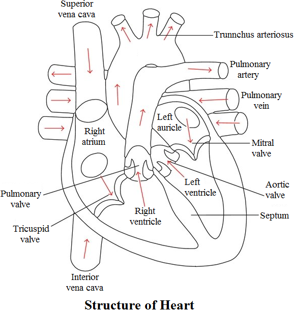
View full question & answer→The sequential events in the heart which are repeated cyclically is called cardiac cycle and it consists of systole (contraction) and diastole (relaxation) of both the atria and ventricles. The duration of a cardiac cycle is 0.8 seconds. Periods of cardiac cycle are atrial systole (0.1 second), ventricular systole (0.3 second) and complete cardiac diastole (0.4 second). The amount of blood pumped by heart per minute is called cardiac output. It is calculated by multiplying stroke volume (volume of blood pumped by each ventricle per minute) with heart rate (number of beats per minute). The heart of normal person beats 72 times per minute and pumps out about 70mL of blood per beat. Therefore, cardiac output averages 5000mL or 5 litres.
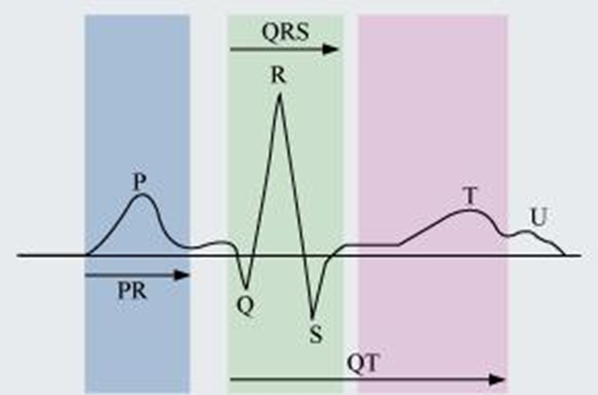 A typical human electrocardiogram has five waves – P, Q, R, S, and T. The P, R, and T-waves are above the base line and are known as positive waves. The Q and S-waves are below the base line and are known as negative waves. The P-wave is of atrial origin, while the Q, R, S, and T-waves are of ventricular origin.