Questions

4 Marks Questions

Take a timed test

22 questions · self-marked practice — reveal the answer and mark yourself.

Question 14 Marks
(i) Write the name of the disease occurring as a result of point mutation.
(ii) Explain the lac operon.
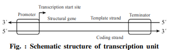
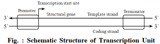
(iii) Draw a labelled diagram of the lac operon.
Answer
(i) The name of the disease occurring as a result of point mutation is Sickle Cell Anaemia.
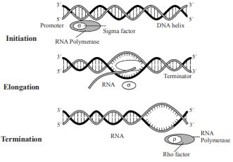
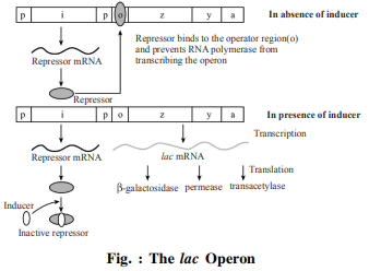
(ii) Lac Operon : It was Jacob and Monod who gave complete elucidation of the Lac operon.
In Lac operon (here lac means lactose) A polycistronic structural gene is regulated by a common promoted and regulator genes. This type of arrangement is very common in bacteria and is known as operon. For example : val-operon, trp-operon, hisoperon, ara-operon etc.
E. Coli bacteria inhabiting in human's intestine derives energy by catabolism of lactose. Jacob and Monod reported that it has a group of three genes in it's DNA which are associated with the synthesis of three enzymes needed for lactose catabolism.
When lactose is present in medium these genes become active but become inactive in absence of lactose Jacob and Monod proposed the operon concept for regulation of these genes activity.
According to this concept, the regulation of gene activity is made at transcription level by induction or repression.
Three enzymes are needed for catabolism and energy production from lactose. These enzymes are
(i) $\beta$galactosidase,
(ii) Permease,
(iii) Transacetylase.
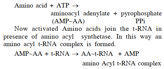
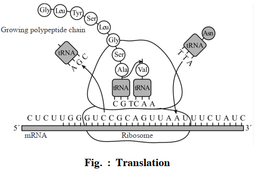
These enzymes are synthesized from translation of poly cistronic m-RNA. The synthesis of this polycistronic mRNA takes place by transcription of three structural genes located in lac operon.
These structural genes or cistrons are—(i) Cis-Z, (ii) Cis-Y and (iii) Cis-A. These are placed adjacently and are mutually coordinated. The activity of structural genes are controlled by regulator gene, operator gene and promoter gene. Cis Z codes for $\beta$ galactosidase which hydrolyses the lactose sugar into glucose and galactose. Cis-Y, codes for permease that increases the permeability of cell for $\beta$ galactosidase. Cis A codes for transacetylase. Therefore the product of all three genes are essential for metabolism of lactose in lac operon.
(iii) Labelled Diagram of the lac operon :
Image
View full question & answer
Question 24 Marks
(i) Define translation.
(ii) Explain the translation process.
(iii) Draw a labelled diagram of the translation.
Answer
self
View full question & answer
Question 34 Marks
Define transcription. Write the names of the parts of the transcription unit in DNA. Draw a schematic diagram of the transcription unit.
Answer
Transcription : Genetic information are transferred from DNA to RNA. The process of synthesis of RNA from DNA is called Transcription.
There are three main components of transcriptional unit in DNA.
(i) Promoter,
(ii) Structural gene,
(iii) Terminator.
Schematic Diagram of the Transcription Unit :
Image
View full question & answer
Question 44 Marks
How did Hershey and Chase differentiate between DNA and protein in their experiment while proving that DNA is the genetic material?
Answer
A.D. Harshey and M.J. Chase conducted experiments on $T _2$ viruses that infect bacteria called bacteriophage and concluded that DNA was the genetic material. Only the DNA of bacteriophage infecting the host enters the bacterial cell which transport the genetic information resulting into the formation of new phase particles.
Hershey and Chase based their experiment on the fact that DNA but not the protein contains phosphorus and similarly Sulfur is present in protein but not in DNA. They then incorporated radioactive isotopes of phosphorus P-32 into phage DNA and that of S-35 into proteins of a separate phage culture.
These phage types were used independently to infect the bacterium E. coli. After some time this mixture was agitated on a blender to separate the empty phage caprices or ghost off the bacterial cells and the two were separated by centrifugation. Hershey and Chase showed that when P-32 was used, all radioactivity was associated with bacterial cells and if followed appeared in the progeny phage. However when S-35 was used, all radioactivity material was limited to phage ghosts. The conclusion was simple and straight forward DNA alone is able to impart all the characteristics to the phage progeny and protein did not play any role in this process.
View full question & answer
Question 54 Marks
Briefly describe the following: Translation
Answer
Translation: The formation of a poly-peptide chain from m-RNA is known as Translation. All three types of RNAs named m-RNA, r-RNA and t-RNA participate in this process. The process of translation takes place on ribosome. The sequence of three nitrogen bases present in 5-3 direction on m-RNA is called genetic code. This code determines the sequence of amino acids in a poly-peptide chain. t-RNA has anticodon which brings specific amino acid from cytoplasm tom-RNA and this entire process requires energy provided by ATP.
The process of translation is completed in following steps:
Activation of amino acids: All the 20 types of amino acids are present in cytoplasm in inactive state but before joining to t-RNA, amino acids become active to react with ATP in presence of $Mg ^{++}$ and amino acyl t-RNA synthetase. The activated amino acids are called amino acyl adenylate.
Image
Involvement of Ribosome: Ribosomes are composed of Protein and r-RNA. These ribosomes are made of two dissimilar (smaller and bigger) subunits which join together at the time of protein synthesis along with m RNA. Many ribosomes unite together forming Polyribosome which takes part in this process.
Initiation of poly-peptide chain formation: Each poly-peptide chain always begins with meithionine amino acid. This amino acid is coded by initiating codon AUG. The t-RNA possessing this amino acid has anti codon UAC. Therefore t-RNA with anti-codon UAC and activated methionine amino acid joins the m-RNA with codon AUG and initiates the synthesis of poly-peptide chain. This process requires the GTP bound with bigger sub-unit of ribosome and initiation factors like $IF _1$, $IF _2$ and $IF _3$ During translation, m RNA contains the sequences which has initiation codon AUG at one end and termination codon at other end. Ribosome binds at 5' end of m RNA and translates codons in m RNA on moving towards 3' end. m-RNA has some additional sequences which are not translated such sequences are said to be untranslated region or UTR. These UTR are very essential for effective translation and are situated before initiation. codon at 5' and after terms nation codon at 3'.
Formation and elongation of poly-peptide chain:
Amino acid in poly-peptide chain are joined together by peptide linkage. This linkage is formed between carboxyl group (-COOH) of one amino acid and an amine group $\left(- NH _2\right)$of next amino acid. The carboxyl group of chain initiating amino acid meithionine remains free to form linkage with - $- NH _2$group of next amino acid.
Now the next activated t-RNA with an appropriate anticodon (that is complementary to codon of m RNA) and amino acid combines with the code of m RNA at A-site. Peptide-linkage is formed between successive amino acids in presence of peptidyl transferase m-RNA moves forward at P site/D-site and A site is occupied by new codon. Now next t-RNA molecule with amino acid reaches the A Site and repeats the same process resulting in elongation of poly peptide chain. The shifting of m RNA from A-site to P-site is called translocation.
Image
Termination of poly peptide chain: The elongation continues till the whole m RNA is translated and a signal in the form of termination codon is reached for which t-RNA has no anticodon such codon are called nonsense codon or stop codon or termination cordons e.g. UAA, UAG and UGA Releasing factors $R _2$ and $R _3$ are helpful in this process which makes polypeptide chain to be free. After becoming free, polypeptide chain undergoes some changes. Enzyme formylase separates the former group of first-amino acid methionine. Some amino acid can be removed from beginning or ending side of chain. After this chain becomes folded to form secondary or tertiary structure. This entire process takes energy from GTP.

View full question & answer
Question 64 Marks
Briefly describe the following: Polymorphism
Answer
Polymorphism: Polymorphism (variation at genetic level) arises due to mutations. New mutations may arise in an individual either in somatic cells or in the germ cells. If a germ cell mutation does not seriously impair individual's ability to have offspring who can transmit the mutation, it can spread to the other members of population (through sexual reproduction). Allelic sequence variation has traditionally been described as a DNA polymorphism if more than one variant (allele) at a locus occurs in human population with a frequency greater than 0.01. In simple terms, if an inheritable mutation is observed in a population at high frequency, it is referred to as DNA polymorphism. The probability of such variation to be observed in non coding DNA sequence would be higher as mutations in these sequences may not have any immediate effect/impact in an individual's reproductive ability. These mutations keep on accumulating generation after generation, and form one of the basis of variability/polymorphism. There is a variety of different types of polymorphisms ranging from single nucleotide change to very large scale changes. For evolution and speciation, such polymorphisms play very important role, and you will study these in details at higher classes.
View full question & answer
Question 74 Marks
Briefly describe the following: Transcription
Answer
Transcription: Genetic informations are transferred from DNA to RNA. The process of synthesis of RNA from DNA is called transcription. DNA directs the synthesis of proteins. This DNA is found in nucleotide of prokaryotic cells and nucleus of eukaryotic cells. DNA molecules never leave their place and therefore DNA molecules code the messages of protein synthesis in mRNA which carries the message from nucleus to cytoplasm over ribosome therefore the synthesis of RNA on DNA template is called transcription. When mRNA is synthesized on DNA template, it contains Uracil in place of Thymine. This synthesis occurs in presence of enzyme RNA polymerase.
A segment of DNA that transcripts the RNA, is called Transcriptional unit. This unit may be like a gene or may have many continuous genes. There are three main components of transcriptional unit in DNA:
(1) Promoter,
(2) Structural gene,
(3) Terminator
DNA dependent RNA polymerase catalyses the polymerization in only one direction i.e. 5'→3'. The strand with 3'→5' polarity serves as template and is therefore, called template strand.
Image

The promoter and terminator flank the structural gene in a transcription unit. The promoter is said to be located towards S'-end (upstream) of the structural gene (the reference is made with respect to the polarity of coding strand). It is a DNA sequence that provides binding site for RNA polymerase, and it is the presence of a promoter in a transcription unit that also defines the template and coding strands. The terminator is located towards 3'-end (downstream) of the coding strand and it usually defines the end of the process of transcription. There are additional regulatory sequences that may be present further upstream or downstream to the promoter.

View full question & answer
Question 84 Marks
What is DNA fingerprinting? Mention its application.
Answer
Among human being, 99.9 percent of base sequence is the same, yet there is a difference in the DNA sequences of any two individuals. The difference in these sequences of DNA make every individuals unique in their phenotypic appearance. The difference in these sequences of DNA can easily be determined by fingerprinting. If one aims to find out genetic differences between two individuals or among individuals of a population, DNA fingerprinting is a very quick way to compare the DNA sequences of these individuals.
In DNA fingerprinting, differences existed in some specific regions/of DNA sequences are identified that are known as Satellite DNA. In DNA fingerprinting. the differences of this satellite DNA is determined. The satellite DNA contains small stretches of base pairs repeated many times, it is therefore, also called repetitive DNA. These repetitive DNA can easily be separated from bulk of genomic DNA by caesium chloride density ultra-centrifugation method in the form of bands. The major peaks are obtained for bulk DNA and lower peaks for satellite/repetitive DNA. This technique was developed by Alec Jeffrey. This technique needs the use of DNA probe. Alec Jeffrey used satellite DNA as probe-as it shows very high degree of polymorphism.
Applications: This technique is now used to:
(1) Identify the criminals in forensic laboratories.
(2) Determine paternity, that is, who the true biological father or mother is of a child.
(3) Verify whether a hopeful immigrant is, as, he or she claims, really a close relative of already an established resident.
(4) identify racial groups to rewrite biological evolution.
View full question & answer
Question 94 Marks
In the medium where E. coli was growing. lactose was added, which induced the lac operon. Then, why does lac operon shut down some time after addition of lactose in the medium.
Answer
It was Jacob and Monad who gave complete explanation of the Lac operon. In Lac operon (here lac means lactose) A poly-cistronic structural gene is regulated by a common promoted and regulator genes. This type of arrangement is very common in bacteria and is known as operon. E Coli bacteria inhabiting in human's intestine derives energy by catabolism of lactose. Jacob and Monad reported that it has a group of three genes in its DNA which are associated with the synthesis of three enzymes needed for lactose catabolism.
When lactose is present in medium these genes become active but become inactive in absence of lactose Jacob and Monad proposed the operon concept for regulation of these genes activity. According to this concept, the regulation of gene activity is made at transcription level by induction or repression. Three enzymes are needed for catabolism and energy production from lactose. These enzymes B-galactosidase, permease and transacetylase.
These enzymes are synthesized from translation of polycistronic m-RNA. The synthesis of this polycistronic m-RNA takes place by transcription of three structural genes located in lac operon. These structural genes or cistrons are Cis-Z, Cis-Y and Cis-A These are placed adjacently and are mutually coordinated. The activity of structural genes are controlled by regulator gene, operator gene and promoter gene. Cis z codes for ẞ galactosidase which hydrolyses the lactose sugar into glucose and galactose. CisY, codes for permease that increases the permeability of cell for ẞ galactosidase. Cis A codes for transacetylase. Therefore the product of all three genes are essential for metabolism of lactose in lac operon.
When lactose is present in the medium as inducer, it enters the cell through the action of permease and joins the repressor produced by regulator gene. As a result repressor can not bind to operator. This allows the RNA polymerase assess to the promoter and transcription of a poly cistronic mRNA from structural genes is resulted which translate the required enzymes for metabolism of lactose. Therefore the lac operon is example of the best understood regulatory mechanism of enzyme induction.
Sometimes lactose on binding with repressor alters its structure and switches off the transcription joining the operators. In this situation, lactose is called co-repressor as it activates repressor to make operator inactive.
View full question & answer
Question 104 Marks
(i) Explain the amino acylation process of tRNA and describe its role in the translation process.
(ii) How does the translation process start in prokaryotes (infinite nuclei)? Describe.
(iii) Where are the untranslated regions located in m-RNA and why?
Answer
(i) All the 20 types of amino acids are present in cytoplasm in inactive state but before joining to t-RNA, Amino acids become active to react with ATP in presence of $Mg ^{++}$and amino acyl t-RNA synthetase. The activated amino acids are called amino acyl adenylate.
Amino acid + ATP $\rightarrow$ aminoacyl adenylate (AMP AA) + pyrophosphate PPi
Now activated amino acids join the t-RNA in presence of amino acyl synthetase. In this way an amino acyl t-RNA complex is formed. This process is called charging of tRNA or amino-acylation of tRNA.
$\begin{aligned} \text { AMP AA +t-RNA } & \rightarrow \begin{array}{l}\text { AA t-RNA }+ \text { AMP } \\ \text { amino Acyl t-RNA complex }\end{array}\end{aligned}$
When two such charged t-RNAs are brought close enough the formation of peptide bond between the corresponding amino acids would be favoured energetically. The presence of a catalyst would enhance the rate of peptide bond formation.
(ii) Initiation of translation in prokaryotes : Each poly-peptide chain always begins with meithionine amino acid. This amino acid is coded by initiating codon AUG. The t-RNA possessing this amino acid has anti codon UAC. Therefore t-RNA with anti-codon UAC and activated meithionine amino acid joins the m-RNA with codon AUG and initiates the synthesis of poly-peptide chain. This process requires the GTP bound with bigger sub-unit of ribosome and initiation factors like $IF _1, IF _2$ and $IF _3$.
(iii) m-RNA has some untranslated region or UTR. These UTR are very essential for effective translation and are situated before initiation codon at 5' and after terms nation codon at 3'.
View full question & answer
Question 114 Marks
What do you mean by transcription? Explain the transcription process with a well labelled
diagram.###What is transcription? Explain the transcription process in bacteria by making a labelled diagram.###Explain the transcription process in bacteria and make a diagram.
Answer
In transcription, hereditary informations are transferred from DNA to RNA. During this process, ribo-nucleotides join at one strand of DNA forming the unstable DNA-RNA hybrid. After some time, complementary chain of RNA is separated. This RNA chain has uracil in place of thymine. This process occurs in presence of RNA polymerase enzyme. 
Transcription in Prokaryotes :
  • In prokaryotes, polycistronic and continuous structural genes are found.
  • In bacteria, there are three major types of RNAs : mRNA (messenger RNA), tRNA (transfer RNA), and rRNA (ribosomal RNA). There is single DNAdependent RNA polymerase that catalyses transcription of all types of RNA in bacteria. All three RNAs are needed to synthesise a protein in a cell. The mRNA provides the template, tRNA brings aminoacids and reads the genetic code, and rRNAs play structural and catalytic role during translation.
  • RNA polymerase binds to promoter and initiates transcription (Initiation). It uses nucleoside triphosphates as substrate. This enzyme has two parts :
    (i) Core enzyme
    (ii) Sigma factor $(\sigma)$
  • Core of enzyme is made by four polypeptide chains :
    (i) Two alpha chains : These polypeptides bind with promotor DNA.
    (ii) First beta chain : It serves to bind the nucleotides used for RNA elongation.
    (iii) Second beta chain : It binds with template DNA helping in transcription. 
  • Sigma factor associates with core enzyme make it active. It recognizes the promoter site on DNA and binds the RNA polymerase at promoter site and initiates transcription actively. 
Image
Fig. : Process of Transcription in Bacteria 
  • As sigma associated core RNA polymerase binds at promoter site, DNA helix unzips from this region and two strands separate from each other. Now one strand acts as template and takes part in transcribing the m-RNA.
  • The RNA polymerase is only capable of catalysing the process of elongation. It associates transiently with initiation-factor $(\sigma)$ and termination-factor ( $\rho$ ) to initiate and terminate the transcription, respectively. Association with these factors alter the specificity of the RNA polymerase to either initiate or terminate.
  • In bacteria, since the mRNA does not require any processing to become active, and also since transcription and translation take place in the same compartment (there is no separation of cytosol and nucleus in bacteria), many times the translation can begin much before the mRNA is fully transcribed. Consequently, the transcription and translation can be coupled in bacteria.
View full question & answer
Question 124 Marks
Explain the difference between prokaryotic and eukaryotic DNA.
Answer
Difference between Prokaryotic and Eukaryotic DNA :
S.No.Prokaryotic DNAEukaryotic DNA
1It is found in cytoplasm, mitochondria and chloroplast.It is found in nucleus.
2Much less in amount, less than 0.1 Pg.Much more (> 1 Pg) in amount than prokaryotic DNA.
3It is usually circular, double stranded.It is usually linear, double stranded.
4It is naked, without histone.It is coated with histone.
5It can code for fewer proteins.It can code for far more proteins.
6Non-coding introns are absent.DNA is mosaic of exons (coding regions) and introns (noncoding region).
7G : C contents are more than A : T.A : T contents are more than G : C.
8On beating it is denatured as a tangled mass.It is denatured into 2 separate strands.
9It exists singly.It exists in pairs.
10Repeated sequences absent.Present.
11Very little part of DNA is functionless.Greater part of DNA is noncistronic and functionless.
View full question & answer
Question 134 Marks
What is called nucleosome? Explain the packaging of DNA coil. Draw a labelled diagram of a nucleosome.
Answer
In eukaryotes, this structure is comparatively more complex. Packaging of DNA in chromosome was explained by R.D. Korenberg and J.O. Thomas in 1974. There is several negatively charged regions in the entire length of DNA where positively charged histone protein is bounded. This composite structure of DNA and protein is known as chromatin. This arrangement of chromatin has been called nucleosome by P. outldet and others. Infect chromatin is made of several spherical beaded units placed periodically at a distance of 11 nanometer and each unit is called nucleoside. Nucleosome is made of octamer of four types of histones. The two molecules each of $H _2 A$, $H _2 B$, and $H _3$ and $H _4$ together form the core which is wrapped around by $13 / 4$ turns of DNA. In this way
Nucleosome $=2$ molecule (each of $H _2 A+ H _2 B+$ $\left.H _3+ H _4\right)+13 / 4$ turns of DNA (140 bp).
DNA joining the two successive nucleoside is known as linker DNA. $H _1$ histone protein is found outside the nucleoside and it is also called linkage histone. There are 60 base pairs in Linker DNA. nucleoside can be isolated by nuclease enzyme. Such six nucleosomes get further condensed and come closer to produce a solenoid structure which represents the secondary structure of chromatin.
Image
This solenoid structure undergoes further coiling to produce tertiary and quartnery structure of chromatin. By such type of coiling, the long DNA molecule gets condensed into a small chromosome that further coil and condense at metaphase stage of cell division to form chromosome. The packaging of chromatin at higher level needs the presence of additional proteins called non histone chromosomal protein. In a typical nucleus some regions of the chromatin are loosely packed and stains light and are referred to as euchromatin. The chromatin that is more densely packed and stains dark are called as heterochromatin.
View full question & answer
Question 144 Marks
Explain the difference between the following :
(i) Nucleoside and nucleotide
(ii) DNA and RNA
(iii) Codon and anticodon
(iv) Transcription and translation
(v) Inductive regulation and inhibitory regulation
(vi) RNA splicing and RNA editing
Answer
(i) Difference between Nucleoside and Nucleotide : 
Nucleoside : The structure formed by the joining of one molecule of sugar with one molecule of nitrogenous base is called nucleoside
Nitrogenous base + sugar = nucleoside. 
Nucleotide : By joining one molecule of phosphoric acid with one molecule of nucleoside, a nucleotide is formed.
Nitrogenous base + sugar + phosphoric acid = nucleotide
(ii) Differences between DNA and RNA :
S.NoCharacterDNARNA
1LocationIt usually occurs in nucleus and some cell organelles like mitochondria and plastid.It usually occurs both in nucleus and cytoplasm.
2NatureGenetic Material.Except some viruses, It is non-genetic In nature.
3No. of StrandUsually double stranded with exception $\phi 174$ bacteriophageUsually single stranded.
4Pentose SugarDeoxyriboseRibose
5Pyrimidine N-BasesCytosine and thymineCytosine and thymine
6Purine and pyrimidine amountEqual in all DNANever equal in RNA
7Unsual N-BasesVery few or asbsentMany and often present
8Pairing of N-basesIs found along entire lengthOnly found in folded part
9Copier enzymeDNA polymeraseRNA polymerase
(iii) Differences between Codon and Anticodon :
S.NoCodonAnticodon
1It is found in DNA and mRNA.It occurs in t-RNA.
2Codon is complementary to a triplet of template strand.It is complementary to a codon.
3It determines the position of an amino acid in a polypeptide.It helps in bringing a particular amino acid at its proper position during translation.
(iv) Differences between Transcription and Translation :
S.NoTranscriptionTranslation
1It is the synthesis of RNA from DNAIt is the synthesis of polypeptide chain from m-RNA.
2It is cytoplasmic event in prokaryotes and nuclear event in eukaryotes.It is cytoplasmic event both in prokaryotes and eukaryotes
3Template is antisense strand of DNA.Template is m-RNA.
4The raw materials are ribonucleotides.The raw materials are 20 types of aminoacids.
5It involves synthesis of all three types of RNA.All three types of RNA take part in synthesis of polypeptide chain.
6It requires RNA polymerases and some transcriptions factors.It requires ribosomes, initiation factors and elongation factors and releasing factors
7RNA polymerases moves over the template.Ribosome moves over the template.
(v) Differences between inductive regulation and inhibitory regulation :
S.NoInductionRepression
1It turns the operon on.It turns the operon off.
2It starts transcription and protein synthesis.It stops transcription and protein synthesis.
3It is caused by a new metabolite which needs enzymes to get metabolised.It is caused by an excess of existing metabolite.
4It operates in a catabolic pathway.It operates in an anabolic pathway.
5Repressor is prevented by the inducer from joining the operator gene.Aporepressor is enabled by a corepressor to join the operator gene.
 (vi) Differences between RNA Splicing and RNA Editing :
S.NoRNA SplicingRNA Editing
1It is the process of removal of introns fromhn-RNA.It involves modification in structure of different types of RNA
2It occurs before translation.It occurs during protein synthesis.
3It occurs in eukaryotes.It occurs both in prokaryotes and eukaryotes


View full question & answer
Question 154 Marks
Write a short note on the following :
(i) Chargaff's equivalence rule
(ii) Replication in semi-conservative mode
(iii) Clover leaf model
(iv) Spliceosome
(v) Termination Codon
(vi) Reverse transcription
(vii) Polyribosome
(viii) hn-RNA
Answer
(i) Chargaff's equivalence rule : Chargoff obtained DNA from various sources in 1949 and studied it to make rules which are called Chargaff's rule. There are :
1. The total amount of purine is equal to the amount of pyrimidine (A+G =T+C)
2. The ratio of A and T and G and C is equal to one
(ii) Replication in semi-conservative mode : Regarding the replication of DNA, the opinion of semi-conservative replication presented by Meselson and Stahl Hall (1958) is universally accepted. According to this, both the strands of the DNA molecule maintain their existence separately from each other and each strand. They synthesize their complementary strand from the pool of nucleotides available in the cell. In this way, in the newly formed DNA molecule, one strand of the previous DNA molecule is newly synthesized, that is, half the same as before and half new, this is called semi-conserved replication. Meselson and Stahl confirmed this by using the heavy isotope $N ^{15}$ on E. coli bacteria.
(iii) Clover leaf model : Robert Holley presented a model of the structure of t-RNA, which is called clover leaf model. According to the clover leaf model, a polynucleotide chain of RNA gets folded to form five arms. Due to bending, the $5^{\prime}$ and $3^{\prime}$ ends come closer. There is a CCA sequence at the $3^{\prime}$ end of each t-RNA molecule. A specific amino acid is added to it with the help of amino acid synthetase enzyme. There is a $G ^{\prime}$ at the $5^{\prime}$ end. Two arms-(i) T $\psi$ C arm : With its help, t-RNA binds with ribosomes and (ii) D arm or DHU arm : It participates in binding with amino acid synthetase. The sequence of three bases on the loop part below t-RNA is anticodon. This m-RNA base pairs with specific codon.
(iv) Spliceosome : In eukaryotes, the RNA formed after transcription is called hnRNA. RNA is made up of two types of parts, one of which is called intron, which does not contain code, the other part is called exon, which carries the genetic code. Intron is removed from it by the process of RNA splicing. Nuclear components called spliceosome help in this process
(v) Termination Codon : These codes are meant to stop or end the elongation of the polypeptide chain. That is, they do not code for any amino acid. These are UAA, UAG and UGA, they are also called stop signals/nonsense codons.
(vi) Reverse transcription : In 1970, Temin and Baltimore discovered that many tumor-causing viruses contain RNA as their genetic material, from which complementary DNA is formed. This reverse transcription is done by transcriptase. Such viruses are called retroviruses. AIDS disease and HIV virus also come under this.
(vii) Polyribosome : Sometimes during protein synthesis, a group of many ribosomes gather on m-RNA. This is called polyribosome. These ribosomes move over the m-RNA from the 5' end to the 3' end. According to the length of m-RNA, 5 to 20 ribosomes are added one after the other. The ribosome that is near the 5' end has the smallest and the one near the 3' end has the longest peptide chain
(viii) hnRNA : It is called heterogeneous nuclear RNA. In eukaryotes, the RNA formed after transcription of DNA is called hnRNA. It is also called high molecular weight RNA or pre-nuclear RNA or DNAlike RNA. This primary messenger RNA is made up of two types of parts. One of these is called intron. There is no code in it. The second part is called exon which carries the genetic code. Of these, introns are removed by the process of RNA splicing. A nuclear organ called spliceosome helps in this process. After this m-RNA is formed, which takes part in the action of translation.
Image
View full question & answer
Question 164 Marks
Explain the difference between the forward strand and lagging strand of DNA.
Answer
Differences between the Leading Strand and Lagging Strand :
Leading StrandLagging Strand
1. It is a replicated strand of DNA which grows continuously without any gap.1. Lagging strand is a replicated strand of DNA which is formed in short segments called Okazaki, fragments. Its growth is discontinuous.
2. It does not require DNA ligase for its growth.2. DNA-ligase is required for joining Okazaki fragments.
3. The direction of growth of the leading strand is $5^{\prime} \rightarrow 3^{\prime}$.3. The direction of growth of the lagging strand is $3^{\prime} \rightarrow 5^{\prime}$ though in each Okazaki fragment it is $5^{\prime} \rightarrow 3^{\prime}$.
4. Only a single RNA primer is required.4. Starting of each Okazaki fragment requires a new RNA primer.
5. Formation of leading strand is quite rapid.5. Formation of lagging strand is slower.
6. Its template opens in $3^{\prime} \rightarrow 5^{\prime}$ direction.6. Its template opens in $5^{\prime} \rightarrow 3^{\prime}$ direction.
7. Formation of leading strand begins immediately at the beginning of replication.7. Formation of lagging strand begins a bit later than that of leading strand.
View full question & answer
Question 174 Marks
What is genetic code? Write their four characteristics and draw a diagram of t-RNA.
Answer
The information of sequence of amino acids exist in the coded language of DNA which is transcribed in mRNA. During transcription, this genetic information is coded in the sequence of nucleotides in m-RNA and determines the sequence of amino acids in protein molecules. The transmission of genetic information between sequence of nitrogen bases of m-RNA and 20 amino acids is known as genetic code. The credit of discovery of genetic code goes to Nirenberg et. al. (1961).
Of all nucleotides, there are only four types of nitrogen bases (A, G, C and U). These four letters are the complete alphabet of genetic dictionary and the sequence of amino acid in protein always follow the sequence of nitrogen bases. The living cells usually have 20 types of essential amino acid in different types of protein and this specific sequence of amino acid in a specific m-RNA depends on the presence of specific type of nitrogen bases that is A, G, C, U sequence.
The characteristics of genetic code are as follow :
1. The code is always triplet : Each codon of the genetic code dictionary is made by specific sequence of three nitrogen bases in m- RNA.
2. The code is comma less : There is no punctuation or comma between two successive code. The codon is read in m-RNA in a contiguous fashion and when the chain is terminated, the terminating codons occupy that place. They are UAA, UAG and UGA.
3. The code is non-ambiguous : Genetic codon does not show ambiguity i.e. a specific codon codes only for one amino acid although some genetic code have been found to code different amino acids in abnormal conditions.
4. The code is non-overlapping : Since the DNA molecule is a long chain of nucleotides it could be read either in an overlapping or non-overlapping manner. In the non-overlapping code, six nucleotides would code for two amino acids, while in the overlapping code up to four could be coded. In the non-overlapping code, each letter is read only once while in the over-lapping code it would be read three times, each time as a part of a different word.
5. The code is universal : It has become proved that genetic code is universal i.e. the genetic code is valid for all organisms ranging from bacteria to man (Except mitochondria and ciliate protozoa i.e.
(i) In yeast mitochondria UGA codes for tryptophan, although in the nuclear genes UGA is a termination codon.
(ii) In ciliate protozoa (Mycoplasma capricolum) in this genetic code, codon UAA and UAG specify glutamine instead of stop signals
The code is degenerated : There are 64 possible codons in a triplet code, of which 61 have been shown to code amino acids. Since only 20 amino acids take part in protein synthesis it is obvious that there are many more codons than amino acid types. Except for tryptophan and methionine, which have a single codon each, all other amino acids involved in protein synthesis have more than one codon. This is called degeneracy of codons. 
View full question & answer
Question 184 Marks
What is the Human Genome Project? Write the features of the Human Genome Project. Give a diagram representing the Human Genome Project.
Answer
Human Genome Project : Before understanding the concept of Human genome project, it is compulsory to know, what is Genome? The gene structure of an individual is called its, genome. As genes are located on chromosomes therefore I a haploid set of chromosomes is said to be genome. The offsprings relieve chromosomes from their parents, the major portion of these chromosomes is DNA. The segment of DNA that contains the genetic information in the form of genetic code, is called gene.
Therefore, it is the sequence of bases in DNA that determines the genetic make up of an individual. Two individuals differ because of difference in DNA sequences at some places. This study led to the quest of finding out the complete DNA sequence of human genome. In 1990, a project was launched for sequencing of complete human genome. In fact it is a mega project.
Aims of HGP : The important goals of HGP are as follows :
(i) Identify all the approximately 20-25 thousands genes in human DNA
(ii) Determine the sequences of the 3 billion chemical base pairs that make up human DNA. (iii) Store these information in data bases.
(iv) Improve tools/new technique for data analysis.
(v) Transfer related technologies to other sectors such as industries.
(vi) Address the ethical, legal and social issues (Elsi). That may arise from the project.]
Image
This project was a 13 year project and was supported by the U.S. department of energy and the national institute of health. Later Japan, France, Germany, China and other countries of the world also contributed in completion of this project. It was completed in 2003. The study of the project has helped in diagnosis, treatment, and prevention of thousands of human disorders by understanding the variation in DNA of different Individuals. On the basis of knowledge the challenges arising in the direction of health, agriculture, energy production, environmental improvement can be solved by using thin natural abilities of DNA sequence. Information has also been obtained about the sequencing of many nonhuman modal organisms like bacterium, yeast, Drosophila, Plants (rice and Arabidopsis).
Salient Features of Human Genome :
Some of the important salient features of HGP are as follows :
(i) The human genome contains 3164.7 million base pairs.
(ii) There are about 3000 bases in an average gene but they differ in sizes. The largest genes so discovered in human is dystrophin which contains 2.4 million bases.
(iii) The total no. of genes is estimated at 30,000 and almost all (99.9%) nucleotides bases are exactly the same in all people.
(iv) The functions are unknown for over 50 per cent of the discovered genes. (v) Less than 2 percent of the genome codes for proteins.
(vi) Repeated sequences makeup very large portion of the human genome.
(vii) Repeated sequences are the stretches of DNA that ale repeated many times, sometimes hundred to thousand times which are considered not to be associated with direct coding functions but they provide knowledge about morphology, dynamics and development of chromosome.
(viii) Chromosome-I has maximum number of genes (2968) and Chromosome-Y has the minimum genes (231).
(ix) Scientists have reported about 1.4 crore location where single base DNA differences (SNPs-single nucleotide polymorphisms known as ‘snips’) occur in humans. The scientists are using this information to find sequences related to chromosomal diseases and tracing human story of evolution.

View full question & answer
Question 194 Marks
Write the components of nucleotide. Write one important feature of the structure of double helical DNA. Draw a labelled diagram of double helical DNA.
Answer
A molecule of Nitrogen base joins the molecule of pentose sugar. It results into the formation of nucleosides. Similarly when phosphoric acid joins the nucleoside it forms nucleotide. i.e. a nucleotide has three components :
1. Pentose sugar (ribose in RNA and deoxyribose in DNA)
2. A phosphate group
3. Nitrogen base
1. Pentose sugar : Depending on the type of pentose sugar, nucleic acid are of two types :
Nucleic acid with ribose sugar is ribonucleic acid (RNA)
Nucleic acid with deoxyribose is deoxyribonucleic acid (DNA) 
Image

The main difference between the two sugars lies at second carbon atom which has -OH group in ribose sugar and hydrogen atom in deoxyribose sugar. Sugar acts as a link to join the nitrogen base and phosphate group.
2. Phosphate or phosphoric acid : This is the ortho-phosphoric acid that connects the two successive nucleotides. This bond is called as phospho-ester bond which joins the nucleoside at 3' carbon atom of sugar to that of 5' carbon atom of successive nucleosides
3. Nitrogen base :
Pyrimidine :
These include thymine, cytosine and uracil of these DNA has thymine and cytosine while RNA has cytosine and uracil.
Purine : They are Adenine and Guanine in DNA and RNA both.
One important feature of the structure of double helical DNA is that DNA is made of two polynucleotide chains which has a backbone of sugar phosphate with nitrogen bases projected inside. The two chains exhibit anti-parallel polarity which means if the one chain has the polarity in 5' to 3' end direction then other has 3' to 5' end.
Image
View full question & answer
Question 204 Marks
What is the difference between transcription in prokaryotic and eukaryotic cells? Compare.
Answer
Differences between transcription in prokaryotic and eukaryotic cells :
S.No.Prokaryotic TranscriptionEukaryotic Transcription
1It occurs in contact with cytoplasm.It occurs inside the nucleus.
2There is no specific period for its occurrence.Major part of transcription occurs in $G _1$ and $G _2$ phases
3Processing of released RNA occurs in cytoplasm.Processing occurs inside the nucleus.
4It is coupled to translation.Transcription and translation are spacially separate.
5Products of transcription become effective in situ.Products of transcription come out of the nucleus for functioning in cytoplasm.
6There is only one RNA polymerase.There are three types of RNA polymerases.
7RNA polymerase does not have separate transcription factors.Transcription factors are involved in recognition of promoter site.
8An enhancer is usually absent.An enhancer may be present alongwith promoter.
9mRNA is generally polycistronic.mRNA is generally monocistronic.
10Splicing is generally not required.In most of the cases splicing is required for removing intervening sequences.
View full question & answer
Question 214 Marks
Draw a labelled diagram and describe the clover leaf pattern model of t-RNA.
Answer
This is the smallest type of RNA made of only 73-94 nucleotides.
  • Its nucleotides chain is folded at many places this is why it has specific 3D structure.
  • The Clover leaf model of tRNA was given by Robert Holley and his co-workers. According to this model, tRNA has 4 arms in two opposite direction :
1. Accepter arm : It is also known as amino acid arm. This arm has both prime ends i.e. 5' and 3' end of tRNA. The 3' of the chain is characterized by the presence of -CCA nitrogen bases which is also known as amino acid attachment site where as the 5' is characterized as caping end.
2. Anticodon arm : Anticodon arm is just opposite to the Accepter arm. It has a specific sequence made of three nitrogen bases called anticodon. The sequence of anticodon is complementary to codon of mRNA therefore it determines the pairing of tRNA with specific code of mRNA. This is why the anticodon on site of the tRNA is also known as mRNA recognition site.
3. T $\psi \quad$ C loop arm : This arm connects the mRNA with ribosome therefore, it is also called ribosome attachment site.
4. Di-hydroxy uridine arm : This arm provides the site for attachment the amino acyl synthetase enzyme by which specific activated amino acid joins t-RNA.
Image
View full question & answer
Question 224 Marks
Describe the molecular structure of DNA by making a labelled diagram.
View full question & answer