Question
Explain the functioning of kidney.

Answer

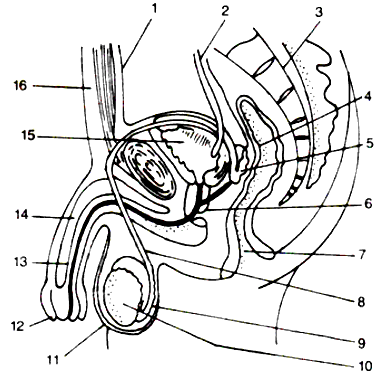
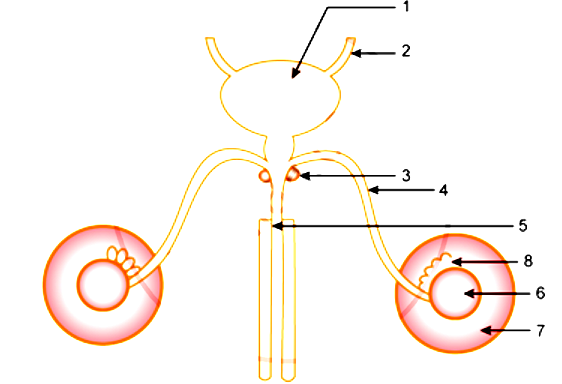
The kidney plays a major role in the formation of urine. The formation of urine takes place in two steps:
(a) Ultrafiltration: The blood flows through the glomerulus under great pressure. This high
pressure causes the liquid part of the blood to filter out from the glomerulus into the renal tubule (ultrafiltration). During ultrafiltration almost all the liquid part of the blood comes out of the glomerulus and passes into the funnel-shaped Bowman’s capsule. The fluid entering the renal tubule is called the glomerular filtrate consisting of water, urea, salts, glucose, and other plasma solutes. The thicker part of the blood left behind in the glomerulus after ultrafiltration, namely, the two kinds of corpuscles, proteins, and other large molecules are carried forward through the efferent arteriole. Thus, the blood proceeding away from the glomerulus is relatively thick.
(b) Reabsorption: The glomerular filtrate entering the renal tubule is an extremely dilute solution containing a lot of usable materials including glucose and some salts such as those of sodium. As the filtrate passes down the tubule, much of the water is reabsorbed together with the usable substances. But their reabsorption is only to the extent that the normal concentration of the blood is not disturbed. This is called selective reabsorption. The fluid which flows through the last part of the tubule is urine.
Certain substances like potassium (K) in the normal course, and a large number of foreign chemicals including drugs like penicillin are passed into the forming urine in tubular wall, and hence it is called tubular secretion.

Need a full question paper?

Generate a complete, print-ready paper with questions like this in minutes — across 16+ boards, with answer keys.

Start Generating Free

Similar questions

The diagram below represents a stage during cell division. Study the same and then answer the question that follow:

a) Name the parts labelled 1, 2, and 3.
b) Identify the above stage and give a reason to support your answer.
c) Mention where in the body this type of cell division occurs.
d) Name the stage prior to this stage and draw a diagram to represent the same.
The diagram given below represents a certain phenomenon which occurs during meiosis. Name and explain the phenomenon by using the terms – homologous chromosomes, chromatids, crossing over.
Given below is a diagram representing a stage during mitotic cell division in an animal cell examine it carefully and answer the questions which follow.

a) Identify the stage. Give one reason in support of your answer.
b) Name the cell organelle that forms the ‘aster’
c) Name the parts labelled 1, 2 and 3.
d) Name the stage that follows the one shown above, how is that stage identified?
e) Mention two differences between mitosis and meiosis with regards to:
(i) The number of daughter cells produced.
The chromosome number in the daughter cells.
List the events taking place in the photo-chemical phase of Photosynthesis.
Given on the right is a diagram which represents the reproductive organs of a man:
(i) Write the names of the parts numbered 1 to 16.
(ii) Give two functions of the part 6.
(iii) Name three components of the fluid which may be found in the tube labeled 2.
(iv) What is the main function of the part labelled 3?
Is ozone depletion related to global warming?
Given below is the outline of the male reproductive system. Name the parts labelled 1 to 8 and state their functions. Also name the corresponding structure of part (4) in the female reproductive system.
The below diagram represents the vertical view of the human female reproductive system.

(i) Label the parts indicated by the guidelines 1 to 8.
(ii) How does the uterus prepare for the reception of a zygote?
(iii) What happens to the uterus if fertilization takes place?
(iv) What happens to the uterus if fertilization has failed to take place?
How is movement in plants different from that in animals?
Name the hormones secreted by the following glands:
(i) Anterior pituitary
(ii) Testes
(iii) Ovary
(iv) Adrenal cortex
(v) Pancreas