Question
Given below is a diagram depicting a defect of human eye. Study the same and then answer the questions that follows.

Image
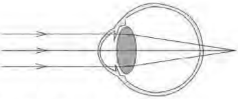
(i) Identify the defect shown in the diagram.
(ii) Mention the cause of this defect.
(iii) Which lens is used to correct this defect?

Answer

(i) Hypermetropia
(ii) The causes of hypermetropia includes: the focal length of the eye lens is too long, or the eyeball has become too small.
(iii) This defect can be corrected by using a convex lens.

Need a full question paper?

Generate a complete, print-ready paper with questions like this in minutes — across 16+ boards, with answer keys.

Start Generating Free

Similar questions

Differentiate between
Nervous control and Hormonal control
Given below is a simple diagram of the circulation of blood in a mammal showing the main blood vessels, the heart, lungs, and body tissues. The blood vessel labeled 6 contains deoxygenated blood and 2 the valve leading to it has three semi-lunar pockets.

(i) Name the blood vessels of organs marked by numbers 1 to 8.
(ii) What do you mean by the term ‘double circulation’ of blood in mammals?
(iii) What is diastole?
Draw neat and labelled diagrams of a myelinated neuron
The diagram given below represents the human heart in one phase of its activity. Study the same and then answer the questions that follow:

(i) Name the phase.
(ii) Which parts of the heart are contracting in this phase ? Give a reason to support your answer.
(iii) Name the part numbered 1 to 6.
(iv) What type of blood flows through the parts marked ‘1’ and ‘2’?
(v) How many valves are closed in this phase?
The diagram given below represents a section of the human heart. Answer the questions that follow:

Image
(i) Which parts of heart are in the diastolic phase? Give a reason to support your answer.
(ii) Label the parts numbered as 1 and 2 in the diagram. What type of blood flows through them?
(iii) What causes the heart sounds 'LUBB' and 'DUBB'?
Differentiate between
Diabetes mellitus and Diabetes insipidus
Differentiate between sensory nerve and motor nerve.
The figure given below represents an experimental set up with a weighing machine to demonstrate a particular process in plants. The experimental set up was placed in bright sunlight. Study the diagram and answer the following questions:

(i) Name the process intended for the study.
(ii) Define the above-mentioned process.
(iii) When the weight of the test tube (A & B) is taken before and after the experiment, what is observed ? Give reasons to justify your observation in A & B.
(iv) What is the purpose of keeping the test tube B in the experimental setup?
In the figure below ‘A’ shows a cell in the normal state and ‘B’ shows the same cell after leaving it in a certain solution for a few minutes.


(i) Describe the change which has occurred in the cell as seen in B.
(ii) Give the technical term for the condition of the cell as reached in B and as it was in A.
(iii) Define the process which led to this condition.
(iv) What was the solution-isotonic, hypotonic or hypertonic, in which the cell was kept?
(v) How can the cell in B, be brought back to its original condition?
(vi) Name the parts numbered 1 to 3.
Differentiate: Endocrine gland and Exocrine gland.