Questions

5 Marks Questions

Take a timed test

40 questions · self-marked practice — reveal the answer and mark yourself.

Question 15 Marks
Describe various types of epithelial tissues with the help of labelled diagrams.
Answer
Epithelial tissue lines the surface of a body and forms a protective covering. Epithelium cells are packed tightly together with little intercellular matrix. Epithelial tissue in the body is of two types:
  1. Simple epithelium: It consists of a single layer of cells where cells are in direct contact with the basement membrane. It is further sub-divided into the following types:
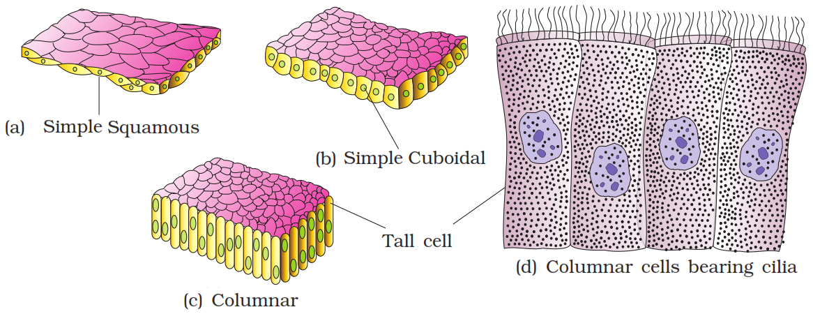
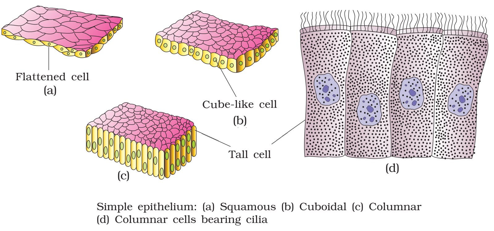
  1. Simple squamous epithelium: It consists of a single layer of flat cells with irregular boundaries. It is foundin the walls of the blood vessels and in the lining of alveoli.
  2. Simple cuboidal epithelium: It consists of a single layer of cube-like cells. It is present in regions where secretion and absorption of substances takes place such as the proximal convoluted tubule region of the nephron.
  3. Simple columnar epithelium: It consists of a single layer of tall, slender cells with their nuclei present at the base of the cells. They may bear micro-villi on the free surfaces. Columnar epithelium forms the lining of the stomach and intestines, and is involved in the function of secretion and absorption.
  4. Ciliated epithelium: It consists of columnar or cuboidal cells with cilia on their free surfaces. They are present in bronchioles and oviducts from where they direct mucus and eggs in specific directions.
  5. Glandular epithelium: It consists of columnar or cuboidal cells involved in the secretion of substances. Glands are of two types, unicellular glands (goblet cells of the alimentary canal) and multicellular glands (salivary glands). They can be classified as exocrine (ductless glands) and endocrine glands (duct glands) by the method through which they release enzymes.
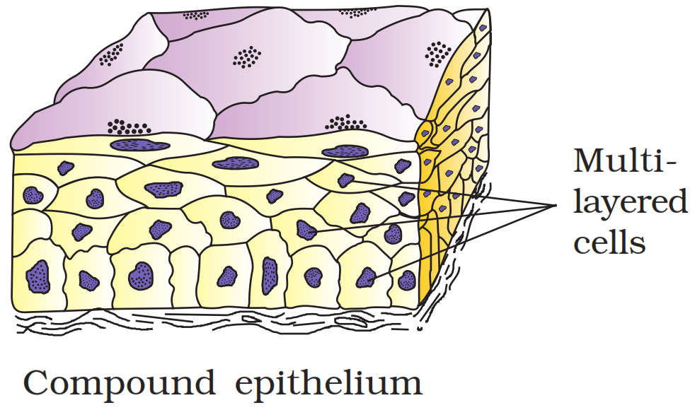
  1. Compound epithelium: It consists of many layers of cells. It is involved mainly in the function of providing protection and has a limited role in secretion and absorption. Examples of compound epithelium include the dry surface of the skin or moist inner lining of the buccal cavity, pharynx, pancreatic ducts, and the inner lining of ducts of salivary glands.
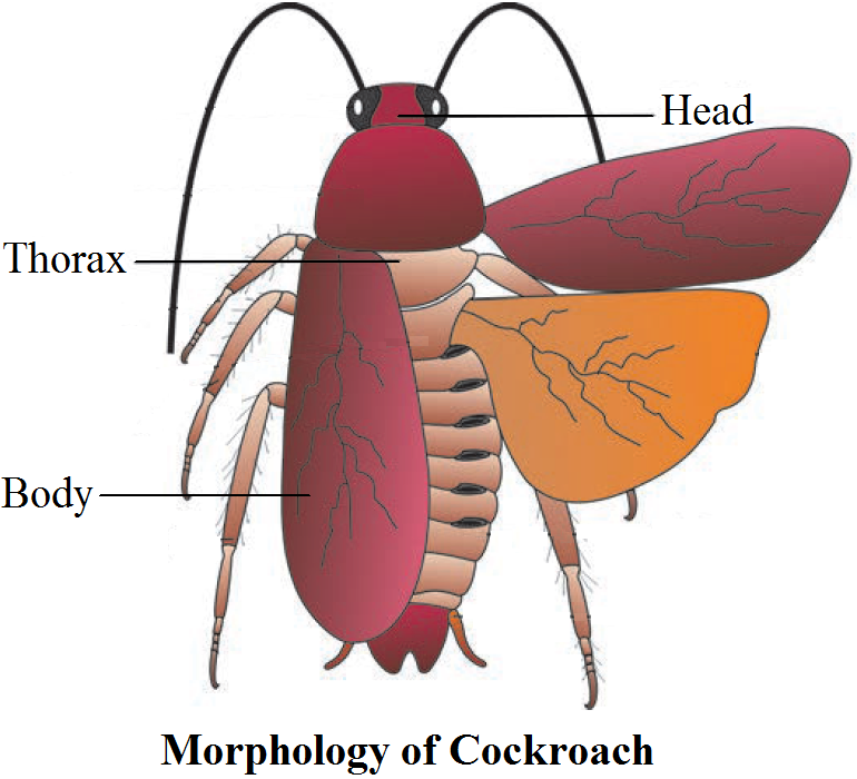
View full question & answer
Question 45 Marks
Match the terms in column I with those in column II:
 
Column I
 
Column II
(a)
Compound epithelium
(i)
Alimentary canal
(b)
Compound eye
(ii)
Cockroach
(c)
Septal nephridia
(iii)
Skin
(d)
Open circulatory system
(iv)
Mosaic vision
(e)
Typhlosole
(v)
Earthworm
(f)
Osteocytes
(vi)
Phallomere
(g)
Genitalia
(vii)
Bone
Answer
 
Column I
 
Column II
(a)
Compound epithelium
(iii)
Skin
(b)
Compound eye
(iv)
Mosaic vision
(c)
Septal nephridia
(v)
Earthworm
(d)
Open circulatory system
(ii)
Cockroach
(e)
Typhlosole
(i)
Alimentary canal
(f)
Osteocytes
(vii)
Bone
(g)
Genitalia
(vi)
Phallomere
View full question & answer
Question 55 Marks
What are the following and where do you find them in animal body.
  1. Chondriocytes.
  2. Axons.
  3. Ciliated epithelium.
Answer
  1. Chondrocytes: Chondrocytes are the only cells found in cartilage. They are present in spaces called lacunae and they produce and maintain the matrix of cartilage. Bending ability of cartilage is due to chondrocytes. Cartilage is present at tip of nose, pinna of ear, epiglottis etc.
  2. Axon: Axon is one of the processes of neuron, which is the structural and functional unit of nervous system. The part of cyton – n’here axon arises is axon hillock and axon ends in group of branches called terminal arborizations. It conducts impulses away from the cyton. Neurons (nerve cells) are present in brain and spinal cord.
  3. Ciliated epithelium: If the columnar or cuboidal cells bear cilia on their free surface they are called ciliated epithelium. Their function is to move particles or mucus in a specific direction over the epithelium. They are mainly present in the inner surface of hollow organs like bronchioles and Fallopian tube.
View full question & answer
Question 65 Marks
Once Amit's teacher took his class to an educational tour where they saw people manufacturing leather from the skin of different animals by the tanning process. Amit found this an interesting process and asked many questions regarding this. After knowing the process, he argued that it is unethical and illegal to use animals for obtaining leather. The teacher appreciated Amit for his action.
  1. Which features of this tissue are useful to obtain leather?
  2. Name the kind of dense connective tissue involved in this.
  3. Write any two unique features of this tissue.
  4. What values of Amit are reflected from the above discussion?
Answer
  1. Features of dense connective tissue are:
  1. It provides strong attachment between various body organs.
  2. The presence of white fibrous tissue at the joints between the skull bones makes them immovable.
  3. Due to the presence of abundant white fibres, the skin dermis of large mammals yields leather after tanning.
  1. White fibrous connective tissue or dense regular connective tissue.
  2. Features of white fibrous connective tissue are:
  1. Rich in white collagen fibres.
  2. The tissue is very tough and inelastic.
  1. Amit is very kind, sharp minded and posseses good moral and ethical values.
View full question & answer
Question 75 Marks
Explain fertilization and development in pheretima.
Answer
Fertilization and Development in Pheretima:
  • A mutual exchange of sperm occurs between two worms during mating. One worm has to find another worm and they mate juxtaposing opposite gonadal openings exchanging packets of sperms called spermatophores.
  • Mature sperm and egg cells and nutritive fluid are deposited in cocoons produced by the gland cells of clitellum. Fertilisation and development occur within the cocoons which are deposited in soil.
  • The cocoon holds the worm embryos. After about 3 weeks, each cocoon produces two to twenty baby worms with an average of four.
  • Earthworms development is direct, i.e., there is no larva formed.
View full question & answer
Question 85 Marks
Discuss the economic importance of the earthworms. How would their absence affect the environment?
Answer
Earthworms are useful in several ways for humans:
  1. Earthworms make the soil porous by digging burrows in the soil. Hence, they are called friends of the farmers.
  2. The nitrogenous wastes and other waste products of the earthworms form food for plants. This process of increasing fertility of soil by earthworms is called vermicomposting.
  3. Earthworms are used as fish bait for catching fishes.
  4. Some tribals in India use earthworms as medicine to cure jaundice, piles, diarrhoea, bladder stones, gout, etc.
  5. In some countries like China, Japan, Australia and Myanmar, earthworms are used as food.
  6. The worms reduce both acidity and alkalinity of the soil and create optimum conditions for the plant growth.
  7. Earthworms are eaten by frogs, birds, which are useful to man in some ways. Thus, they are an important part of food chain.
  8. These are used in scientific studies and dissected in zoological laboratories for academic studies.
View full question & answer
Question 95 Marks
Using appropriate examples, differentiate between false and true body segmentation.
Answer
DIFFERENCES BETWEEN TRUE AND PSEUDO (false) METAMERISM
  • True metamerism commonly occurs in the arthropods, chordates, and the annelids. While Pseudo-metamerism occurs in the tapeworms etc.
  • In pseudo-metamerism, segmentation of the ectoderm is the base of metamerism in the complete body. While the segmentation of the mesoderm is the base reason of the segmentation in the True metamerism.
  • In the pseudo-metamerism, all the segments work as a single unit and they work independently. While in the true metamerism the units work as different units.
  • True metamerism has the coordination in the segments while the Pseudo-metamerism has zero coordination between the segments.
View full question & answer
Question 105 Marks
Which features distinguish blood from lymph?
Answer
Differences between blood and lymph are given below:
S. No.
Blood
Lymph
1.
It contains plasma, erythrocytes, leucocytes and platelets.
It contains plasma and leucocytes.
2.
The presence of haemoglobin imparts red colour to it.
It is colourless as haemoglobin is absent.
3.
Its plasma contains more protein, calcium and phosphorus as compared to lymph.
Its plasma has fewer protein and less calcium and phosphorus than blood.
4.
Contains moderate amount of $CO_2$ and other metabolic wastes.
Contains excessive amount of $CO_2$ and other metabolic wastes.
View full question & answer
Question 125 Marks
Differentiate between neurons and neuroglia.
Answer
Differences between neurons and neuroglia are:
S. No.
Neurons
Neuroglia
1.
They conduct nerve impulses.
They form packing around the nerve cells in the central nervous system. Some are phagocytic in nature.
2.
They have two types of processes, i.e., axon and dendron.
They have only one type of process.
3.
They form synapse.
They do not form synapse.
4.
They cannot divide.
They can divide.
View full question & answer
Question 135 Marks
Give a detail account on neural tissue.
Answer
The neural tissue is ectodermal in origin. It is specialised to receive stimuli and conducts impulses for controlling and coordinating body functions. It exerts the greatest control over the body's responsiveness to changing conditions. The neural tissue consists of nerve cells and packing cells. The packing cells are called Schwann cells in the peripheral nervous system and neuroglia cells in the central nervous system.
View full question & answer
Question 155 Marks
What are the cellular components of blood?
Answer
Blood is a fluid connective tissue. It is composed of plasma (fluid) and blood cells (corpuscles). Cellular components of blood (blood corpuscles) constitute about $45 \%$ of blood volume. Three types of blood cells are:
i. Erythrocytes or red blood cells: They are most abundant blood cells. Normal RBC count is $5-5.5$ million $/ \mathrm{mm}^3$ in males and 4.5-5 million/ $\mathrm{mm}^3$ in females. RBCs help in transport of gases and maintain blood pH .
ii. Leucocytes or white blood cells: The normal WBC count is $5000-6000 / \mathrm{mm}^3$ of blood. They are involved in immune response of body and act as soldiers and scavangers.
iii. Thrombocytes or blood platelets: There are about 2, 50,000 platelets $/ \mathrm{mm}^3$ of blood. They are involved in blood clotting.
View full question & answer
Question 165 Marks
Mention special features of eye in cockroach.
Answer
The eyes in cockroach are large, sessile, paired, bean-shaped and present on either side of head. These are compound in nature. Each compound eye consists of large number of visual elements called ommatidia. Each ommatidium is composed of a diopteric region and reticular (receptor) region. It is capable of producing a separate image of a small part of 'object seen. Thus, the image of the object viewed consists of several pieces and hence known as mosaic image. From the inner end of each ommatidium, fine nerve fibers arise, all of which combine to form one optic nerve connected to the brain.
View full question & answer
Question 175 Marks
Frog is a poikilotherm, exhibits camouflage and undergoes aestivation and hibernation, how are all these beneficial to it?
Answer
Frog is a poikilotherm (cold blooded), i.e., it cannot maintain constant body temperature and it varies with change in environmental temperature. Being cold blooded, frog cannot withstand very cold weather and extreme high temperature and thus buries itself deep in mud during winters and summers, known as hibernation (winter-sleep) and aestivation (summer sleep) respectively. These periods are of little activities and energy required is obtained from fat bodies, or stored glycogen in liver, which have been stored earlier. Camouflage is the ability to change body colour with change in its surroundings and climatic conditions. It helps the animal to escape from predators or to attack its prey.
View full question & answer
Question 195 Marks
Explain the morphology of an earthworm.
Answer
Morphology of Earthworm: Body: Earthworms have long cylindrical body. The body is divided into more than hundred short segments which are similar (metameres about 100-120 in number). Dorsal Surface: The dorsal surface of the body is marked by a dark median mid dorsal line (dorsal blood vessel) along the longitudinal axis of the body.Ventral Surface: The ventral surface is distinguished by the presence of genital openings (pores).

Segments: In a mature worm, segments 14-16 are covered by a prominent dark band of glandular tissue called clitellum. Thus the body is divisible into three prominent regions.
  • Preclitellar.
  • Clitellar.
  • Costclitellar segments.
Buccal Region: Anterior end consists of the mouth and the prostomium, a lobe which serves as a covering for the mouth and as a wedge to force open cracks in the soil into which the earthworm may crawl. The prostomium is sensory in function. The first body segment is called the peristomium (buccal segment) which contains the mouth.Genital Openings: Four pairs of spermathecal apertures are situated on the ventro-lateral sides of the intersegmental grooves, i.e., 5th-9th segments. A single female genital pore is present in the mid-ventral line of 14th segment. A pair of male genital pores are present on the ventro-lateral sides of the 18th segment.
Locomotion: In each body segment, except the first, last and clitellum, there are rows of S-shaped setae, embedded in the epidermal pits in the middle of each segment. Setae can be extended or retracted. Their principal role is in locomotion.
View full question & answer
Question 205 Marks
Explain the digestive system of cockroach with the help of a labelled sketch.
Answer
The digestive system consists of the alimentary canal and digestive glands. The alimentary canal of cockroach is divided into foregut, midgut, and hindgut. The mouth opens into a short tubular pharynx, leading to a narrow tubular passage, the oesophagus. The oesophagus opens into a sac-like-crop, which is used for storing food. The crop is followed by a gizzard or proventriculus. Gizzard consists of six-chitinous plates called teeth which help in grinding food. The entire foregut is lined by the cuticle. A ring of six-eight blind tubular structures called hepatic or gastric caeca is present at the junction of foregut and midgut which secrete digestive juice. At the junction of midgut and hindgut is present another ring of yellow coloured thin filamentous malpighian tubules which help in removal of excretory products from haemolymph. The hindgut is broader than midgut and is differentiated into the ileum, colon, and rectum. The rectum opens out through the anus.
View full question & answer
Question 215 Marks
Explain morphology of cockroach.
Answer
Morphology of Cockroach: The adults of the common species of cockroach, Periplaneta Americana are about 34-53 mm long with wings that extend beyond the tip of the abdomen in males. The body of the cockroach is segmented and divisible into three distinct regions:
  • Head.
  • Thorax.
  • Abdomen.
Exoskeleton: The entire body is covered by a hard chitinous exoskeleton (brown in colour). In each segment, exoskeleton has hardened plates called sclerites (tergites dorsally and sternites ventrally) that are joined to each other by a thin and flexible articular membrane (arthrodial membrane). Head:
  • Head is triangular in shape and lies anteriorly at right angles to the ongitudinal body axis.
  • It is formed by the fusion of six segments and shows great mobility in all directions due to flexible neck.
  • The head capsule bears a pair of compound eyes.
  • A pair of thread like antennae arises from membranous sockets lying in front of eyes. Antennae have sensory receptors that help in monitoring the environment.
Mouth Parts: Anterior end of the head bears appendages forming biting and chewing type of mouth parts. The mouthparts consisting of a labrum (upper lip), a pair of mandibles, a pair of maxillae and a labium (lower lip). A median flexible lobe, acting as tongue (hypopharynx), lies within the cavity enclosed by the mouthparts. Thorax: Thorax consists of three parts
  • Prothorax.
  • Mesothorax.
  • Metathorax.
The head is connected with thorax by a short extension of the prothorax known as the neck. Each thoracic segment bears a pair of walking legs. The first pair of wings arises from mesothorax and the second pair from metathorax. Forewings (mesothoracic) called tegmina are opaque dark and leathery and cover the hind wings when at rest. The hind wings are transparent, membranous and are used in flight. Abdomen:
  • The abdomen in both males and females consists of 10 segments.
  • In females, the 7th sternum is boat shaped and together with the 8th and 9th sterna forms a brood or genital pouch whose anterior part contains female gonopore, spermathecal pores and collateral glands.
  • In males, genital pouch or chamber lies at the hind end of abdomen bounded dorsally by 9th and 10th terga and ventrally by the 9th sternum. It contains dorsal anus, ventral male genital pore and gonapophysis. Males bear a pair of short, threadlike anal styles which are absent in females.
  • In both sexes, the 10th segment bears a pair of jointed filamentous structures called anal cerci.
View full question & answer
Question 225 Marks
In which segment does the following structures lies in the earthworm's body?
  1. Spermathecae.
  2. Pharynx.
  3. Gizzard.
  4. Intestine.
  5. Septal nephridia.
  6. Ovary.
  7. Testes.
  8. Typhlosole.
  9. Lateral heart.
  10. Pharyngeal nephridia.
Answer
  1. Spermathecae- 6th, 7th, 8th, 9th.
  2. Pharynx- 4th.
  3. Gizzard- 8th- 9th.
  4. Intestine- 15th to last.
  5. Septal nephridia- 15th to last.
  6. Ovary- 13th.
  7. Testes- 10th, 11th.
  8. Typhlosole- 26th- 95th
  9. Lateral heart- 7th, 9th
  10. Pharyngeal nephridia- 4th- 6th 55.
View full question & answer
Question 235 Marks
Describe the circulatory system of frog.
Answer
The circulatory system of frog is well-developed and is of closed type. It also has a lymphatic system. The blood vascular system includes the heart, blood and blood vessels. The lymphatic system consists of lymph, lymph channels and lymph nodes.Heart:
It is a muscular structure situated in the upper part of body cavity. It has three chambers, i.e., two atria (sing, atrium) and one ventricle. The heart is covered by a membrane called pericardium.
There are two accessory chambers in the frog's heart, a tubular truncus arteriosus placed on the ventral side of the right auricle and a triangular sinus venosus present on the dorsal side of the heart.
Sinus venosus receives blood through the major veins called vena cava and opens into right auricle.
The left auricle receives oxygenated blood from the lungs by a common pulmonary vein. The auricles send their blood into ventricle where the blood gets mixed up. The ventricle opens into a sac-like conus arteriosus on the ventral side of the heart.
Special venous connection between liver and intestine as well as the kidney and lower parts of the body are present in frogs.
The former is called hepatic portal system and the latter is called renal portal system.
Blood Vessels:
The blood vessels found in frogs are arteries, arterioles, veins, venules and blood capillaries.
Arteries carry blood from the heart to different body parts. Veins bring blood from different body parts to the heart. Arteries further divide to form arterioles. The arterioles branch out to form capillaries, which further unite to form venules. The venules thus, join to form veins (which have valves to prevent backflow of the blood).
Blood:
The blood is composed of plasma and blood cells. The blood cells are RBCs (Red Blood Cells) or erythrocytes, WBCs (White Blood Cells) or leucocytes and platelets. RBCs are nucleated and contain red coloured pigment namely haemoglobin.
View full question & answer
Question 245 Marks
Write short notes on following:
  1. Malpighian tubules.
  2. Excretion in Frogs.
  3. Economic importance of cockroach.
  4. Glandular Epithelium.
  5. Neuron.
Answer
  1. Malpighian Tubules: The system consists of branching tubules extending from the alimentary canal that absorbs solutes, water, and wastes from the surrounding hemolymph. The wastes then are released from the organism in the form of solid nitrogenous compounds. Malpighian tubules are found in some members of the phylum Arthropoda.
  2. Excretion in Frogs: Frogs have well developed system composed of a pair of kidneys. Kidneys contain many filtering units called nephrons. These nephrons filter waste prouct from blood. The waste then goes to collecting tubules and finally to urinary bladder through ureters. Frogs are ureotelic animals.
  3. Economic Importance of Cockroach: Some species of cockroaches live in human dwellings. They contaminate human food items while they search for food. They can be a major source of nuisance as well as of some of the diseases. Other than them most of the species of cockroaches have little significance for humans.
  4. Glandular Epithelium: Some of the columnar or cuboidal cells get specialized for secretion and are called glandular epithelium. They are mainly of two types:
  • Unicellular, consisting of isolated glandular cells (goblet cells of the alimentary canal).
  • Multicellular, consisting of cluster of cells (salivary gland).
On the basis of the mode of pouring of their secretions, glands are divided into two categories namely exocrine and endocrine glands. Exocrine glands secrete mucus, saliva, earwax, oil, milk, digestive enzymes and other cell products. These products are released through ducts or tubes. In contrast, endocrine glands do not have ducts. Their products, called hormones, are secreted directly into the fluid bathing the gland.
  1. Neurons: the unit of neural system are excitable cells.
Structure: A neuron contains a cell body with nucleus and a tail called axon. On the main cell body there are many hair like structures called dendrites. The axon is covered with myelin sheath which gives insulation.
When a neuron is suitably stimulated, an electrical disturbance is generated which swiftly travels along its plasma membrane. Arrival of the disturbance at the neuron's endings, or output zone, triggers events that may cause stimulation or inhibition of adjacent neurons and other cells and thereby transmitting neural signals to different parts of the body.
View full question & answer
Question 255 Marks
Explain the alimentary canal of a pheretima with suitable diagram. Explain how the peculiar feeding habit of earthworm is helpful for humans?
Answer
Alimentary Canal: The alimentary canal is a straight tube and runs between first to last segment of the body. A terminal mouth opens into the buccal cavity (1-3 segments) which leads into muscular pharynx. A small narrow tube, oesophagus (5-7 segments), continues into a muscular gizzard (8-9 segments). It helps in grinding the soil particles and decaying leaves, etc. The stomach extends from 9-14 segments.
  • The food of the earthworm is decaying leaves and organic matter mixed with soil. Calciferous glands, present in the stomach, neutralise the humic acid present in humus.
  • Intestine starts from the 15th segment onwards and continues till the last segment.
  • A pair of short and conical intestinal caecae project from the intestine on the 26th segment. The characteristic feature of the intestine between 26-35 segments is the presence of internal median fold of dorsal wall called typhlosole. This increases the effective area of absorption in the intestine.
  • The alimentary canal opens to the exterior by a small rounded aperture called anus. The ingested organic rich soil passes through the digestive tract where digestive enzymes breakdown complex food into smaller absorbable units. These simpler molecules are absorbed through intestinal membranes and are utilised.
Feeding habit and importance for human: The earthworm eats soil and decaying organic materials. Because of muscular pharynx the earthworms grinds everything very finely. Calciferous glands of the digestive system help in composting of organic materials of earthworm's food. Finally the waste product come out which is very good compost fertilizer. Because of this the method h astening composting process with the help of earthworm is known as vermin composting. Otherwise also the earthworm facilitates turning over of the soil layer thereby helping in better aeration of the topsoil.
View full question & answer
Question 265 Marks
Cardiac muscles are contractile muscles present in heart.
  1. What is their structure?
  2. Write their special features.
  3. Find the value in non-tiring nature of cardiac muscles.
Answer
  1. The cardiac muscles are contractile tissues present only in the heart and in the wall of large veins which enter the heart. The cardiac muscle fibres show the characters of both unstriped and striped muscle fibres. Each fibre is a long and cylindrical structure which has a definite sarcolemma. The fibres are uninucleate and the nuclei lie near the centre.
  2. The cardiac muscle fibres have some special features:
  1. These muscle fibres are supplied with both central and autonomic nervous system and are not under the will of the animal.
  2. These fibres never get fatigue.
  3. Blood capillaries penetrate the cardiac muscle fibres, hence they have very rich blood supply.
  4. These fibres have the property of contraction, even when they are isolated from the body temporarily.
  1. Cardiac muscle resists fatigue so well because it's got more mitochondria than skeletal muscle. With so many power plants at its disposal, the heart doesn't need to stop and chill out. It also has a steady supply of blood bringing it oxygen and nutrients.
View full question & answer
Question 275 Marks
Draw the diagrams of mouth parts of cockroach and briefly describe them.
Answer
It is a narrow opening that lies at the base of the preoral cavity. It is bounded by the mouth parts and leads into the pharynx. The mouth parts of cockroach are of biting and chewing type. They also help in swallowing. The mouth parts are attached to the head capsule. The mouth parts include the following structures:
  1. Labrum is also called upper lip that helps in holding food particles during feeding.
  2. Mandibles lie on the sides of the mouth just behind the labrum. The two mandibles work against each other in a horizontal plane to crush and cut the food into pieces.
  3. First maxillae are a pair of maxillae that lie beneath the andibles, one on either side of the head.
  4. Second maxillae or labium is also called lower lip. It is a single structure, but it is formed by the fusion of a pair of second maxillae. It lies behind the mouth and forms a type of lower lip.
A median flexible lobe, acting as tongue (hypopharynx) lies within the cavity enclosed by the mouthparts.
View full question & answer
Question 285 Marks
Classiafy and describe epithelial tissue on the basis of structural modifications of cells.
Answer
Epithelial tissues can be divided into three types on the basis of structural modifications of cells, namely squamous, cuboidal and columnar.
  • Squamous Epithelium: This type of epithelial tissue is made up of a single thin layer of flattened cells. The cells in this tissue have irregular boundaries. This type of epithelium is found in the walls of blood vessels and in air sacs in the lungs. The thin layer makes a diffusion boundary and thus facilitates diffusion of substances.
  • Cuboidal Epithelium: This type of epithelial tissue is composed of a single layer of cuboidal cells. This type of tssue is found in the ducts of glands and in nephron. Secretion and absorption are the main functions of cuboidal epithelium. Columnar.
  • Epithelium: This type of tissue is composed of a single layer of tall and slender cells. The nuclei of cells are located at the base. Microvilli may be present at the top of cells. This type of tissue if found in the lining of stomach and intestine. The main function is secrection and absorption.
  • Ciliated Epithelium: If cilia are present on cuboidal or columnar epithelium, they are called ciliated epithelium. In that case, movement of substances is their main function. Ciliated epithelium is found in bronchioles and fallopian tubes.
  • Glandular Epithelium: When cuboidal or columnar epithelium assume specialized function of secreation, they are called glandular epithelium. These are found in exocrine and endocrine glands.
View full question & answer
Question 295 Marks
  1. Draw a labelled diagram of areolar connective tissue.
  2. Where is it found in human body?
  3. How does it differ from adipose tissue?
Answer
  1.  
  1. It is found beneath the skin.
  2.  
S. No.
Areolar Connective Tissue
Adipose Tissue
1.
It serves as a support framework for epithelium.
It synthesises, stores and metabolises fat.
2.
Adipocytes are absent.
Adipocytes are present.
3.
It is found in almost all parts of the body.
It is found mainly beneath the skin.
4.
Mast cells are Present.
Mast cells are absent.
View full question & answer
Question 305 Marks
With the help of appropriate diagrams explain various kinds of muscular tissues with their functions.
Answer
Muscle Tissue:
  • Each muscle is made of many long, cylindrical fibres arranged in parallel arrays.
  • These fibres are composed of numerous fine fibrils, called myofibrils.
  • Muscle fibres contract (shorten) in response to stimulation, then relax (lengthen) and return to their uncontracted state in a coordinated fashion.
  • Their action moves the body to adjust to the changes in the environment and to maintain the positions of the various parts of the body.
Muscles are of three types:
  • Skeletal.
  • Smooth.
  • Cardiac.
Skeletal or Striated Muscle: Skeletal muscle tissue is closely attached to skeletal bones. Striated or skeletal muscle fibres are bundled together in a parallel fashion. A sheath of tough connective tissue encloses several bundles of muscle fibres. Function: Skeletal muscles are 'voluntary' as their function is under the conscious control. They facilitate articulation of limbs, neck, torso, etc. 
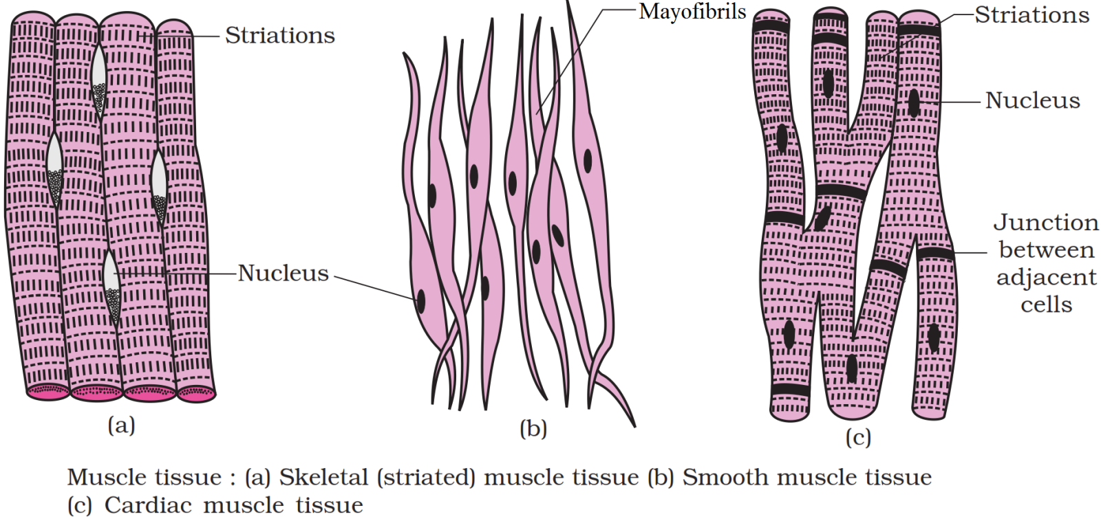
Smooth Muscle: The smooth muscle fibres taper at both ends (fusiform) and do not show striations. Cell junctions hold them together and they are bundled together in a connective tissue sheath. The wall of internal organs such as the blood vessels, stomach and intestine contains this type of muscle tissue. Function: Smooth muscles are 'involuntary' as their functioning cannot be consciously controlled.Cardiac Muscle: Cardiac muscle tissue is a contractile tissue present only in the heart. Cell junctions fuse the plasma membranes of cardiac muscle cells and make them stick together. Communication junctions (intercalated discs) at some fusion points allow the cells to contract as a unit, i.e., when one cell receives a signal to contract, its neighbours are also stimulated to contract.
Function: Facilitate pumping action of heart.
View full question & answer
Question 315 Marks
Explain the reproductive system in cockroach with the help of suitable diagrams.
Answer
Reproductive System: Cockroaches are dioecious and both sexes have well developed reproductive organs. Male Reproductive System:
  • Male reproductive system consists of a pair of testes lying one on each later side in the 4th-6th abdominal segments.
  • From each testis arises a thin vas deferens, which opens into ejaculatory duc through seminal vesicle. The ejaculatory duct opens into male gonopore situate ventral to anus.
  • A characteristic mushroom shaped gland is present in the 6th-7th abdominal segments which functions as an accessory reproductive gland.
  • The external genitalia are represented by male gonapophysis or phallomer (chitinous asymmetrical structures, surrounding the male gonopore).
  • The sperms are stored in the seminal vesicles and are glued together in th form of bundles called spermatophores which are discharged during copulation.
Female Reproductive System:
  • The female reproductive system consists of two large ovaries, lying laterally in the 2nd - 6th abdominal segments. Each ovary is formed of a group of eight ovarian tubules or ovarioles, containing a chain of developing ova.
  • Oviducts of each ovary unite into a single median oviduct (also called vagina which opens into the genital chamber.
  • A pair of spermatheca is present in the 6th segment which opens into thi genital chamber.
Fertilisation:
  • Sperms are transferred through spermatophores.
  • Their fertilised eggs are encased in capsules called oothecae. Ootheca is a dark reddish to blackish brown capsule, about 3/8" (8 mm) long. They are dropped or glued to a suitable surface, usually in a crack or crevice of high relative humidity near a food source.
  • On an average, females produce 9-10 oothecae, each containing 14-16 eggs.
  • The development of P. Americana is paurometabolous, meaning there is development through nymphal stage. The nymphs look very much like adults The nymph grows by moulting about 13 times to reach the adult form. The next to last nymphal stage has wing pads but only adult cockroaches have wings.
View full question & answer
Question 325 Marks
Describe biting and chewing type of mouth parts by taking example of cockroach.
Answer
It is a narrow opening that lies at the base of the preoral cavity. It is bounded by the mouth parts and leads into the pharynx. The mouth parts of cockroach are of biting and chewing type. They also help in swallowing. The mouth parts are attached to the head capsule. The mouth parts include the following structures:
  1. Labrum: Is also called upper lip that helps in holding food particles during feeding.
  2. Mandibles: Lie on the sides of the mouth just behind the labrum. The two mandibles work against each other in a horizontal plane to crush and cut the food into pieces.
  3. First maxillae: Are a pair of maxillae that lie beneath the mandibles, one on either side of the head.
  4. Second maxillae or labium: Is also called lower lip. It is a single structure, but it is formed by the fusion of a pair of second maxillae. It lies behind the mouth and forms a type of lower lip.
A median flexible lobe, acting as tongue (hypopharynx) lies within the cavity enclosed by the mouthparts.
View full question & answer
Question 335 Marks
Differentiate between the striated, non-striated and cardiac muscles.
Answer
  1. Striated Muscles: The striated or skeletal muscles form about 40% of total body weight. These muscles are attached and bring about the movement of the various bones of the skeleton, so are called skeletal muscles. The striated muscles give shape to the body and also release heat during contraction. These muscles have huge supply of nerves and blood vessels. Each striated muscle is a long, narrow, cylindrical, unbranched cell.
The striated muscle fibres are multinucleated or syncytial in nature. The cytoplasm (sarcoplasm) of each fibre has a large number of myofibrils (actin and myosin myofibrils) which are tightly packed. Each myofibril shows dark and light bands of stripes alternating with each other. Hence, they are called as striped muscle fibres.
  1. Non-Striated (Smooth) Muscle: The non-striated muscles are found in the posterior part of oesophagus, stomach, intestine, lungs, urinogenital tract, urinary bladder, blood vessels, iris, ciliary body of eye, dermis of skin, etc.
The non-striated muscle consists of long, narrow, spindle-shaped fibres that are generally shorter than the striated muscle fibres. Their size may range from 20 um (small blood vessels)-500 um (in pregnant uterus).
Each non-striated muscle fibre contains a single oval nucleus in its thick middle part. In the cytoplasm, the myofibrils are arranged longitudinally. They are composed of myosin. There is no sarcolemma, however, the fibre is enclosed by the plasma membrane.
The smooth muscles help in the peristalsis which occurs in the tubular viscera. The autonomic nervous system controls these muscles. Hence, they are not under the control of animal's will.
  1. Cardiac Muscles: The cardiac muscles are contractile tissues present only in the heart and in the wall of large veins which enter the heart. The cardiac muscle fibres show the characters of both unstriped and striped muscle fibres. Each fibre is a long and cylindrical structure which has a definite sarcolemma. The fibres are uninucleate and the nuclei lie near the centre.
The myofibrils have transverse faint dark and light bands which alternate with each other.
View full question & answer
Question 345 Marks
Distinguish between male cockroach and female cockroach.
Answer
Differences between male and female cockroach are:
S. No.
Male Cockroach
Female Cockroach
1.
Abdomen is long and narrow.
Abdomen is short and broad.
2.
All nine sterna are visible.
Only seven sterna are visible.
3.
Anal styles are present.
Anal styles are absent.
4.
$7^{\text {th}}$ tergum covers $8^{\text {th}}$ tergum.
$7^{\text {th}}$ tergum covers $8^{\text {th}}$ and $9^{\text {th}}$ terga.
5.
Brood pouch is absent.
Brood pouch is present.
6.
Antennae are longer in size.
Antennae are smaller in size.
7.
Wings extend beyond the tip of abdomen.
Wings extend upto the end of abdomen.
View full question & answer
Question 355 Marks
Describe mouth parts of the cockroach.
Answer
Mouth Parts of Cockroach. Anterior end of the head bears appendages forming biting and chewing type of mouth parts. The mouth parts consist of following structures:
  1. Labrum (upper lip).
  2. A pair of mandibles.
  3. A pair of maxillae.
  4. Labium (lower lip).
Apart from these a median flexible lobe, acting as tongue (hypopharynx), lies within the cavity enclosed by the mouth parts.
View full question & answer
Question 365 Marks
Describe the circulatory system of cockroach.
Answer
The circulatory system of cockroach is of open type. The blood vessels are poorly developed and blood flows freely in the body cavity which is called haemocoel. Visceral organs located in the haemocoel are bathed in blood (haemolymph). It contains colourless plasma and corpuscles called haemocytes. Each heart chamber opens into next chamber through a ventricular valve. Each chamber has a pair of incurrent pores called ostia (which possess valvular mechanism to pass the blood only from haemocoel to the heart chambers). The heart chambers contract one after the other rapidly. This pushes the blood into the anterior aorta as well as a few lateral or excurrent arteries. From aorta, blood passes into head sinuses and then into perivisceral and perineural sinuses.
View full question & answer
Question 375 Marks
Comment upon the gametic exchange in earthworm during mating.
Answer
Mating in earthworm is a unique process, earthworm is a hermaphrodite organism. Breeding in earthworm takes place during rainy season and begins with copulation soon after maturation of the sperms. The gametic exchange and the physiology reproduction during mating can describe as below
  1. Earthworms are protandrous animal (i.e., maturation of sperm takes place muchearlier then that of ova).
  2. Mating process in earthworm occurs through cross-fertilization.
  3. The mating process involves exchange of sperms between the two worms.
  4. Two individuals from adjacent burrows half emerge out and lie in contact with each other, and opposite gonadal opening exchange the packets of sperms called spermatophores.
  5. During the process, the skin encircling male pore, elevates a little, to form a temporary papilla that fits like a penis into the opposite spermathecal pore to keep it open.
  1. After filling of spermathecal, the copulating worm moves a bit to adjust another pair of spermathecae to face the male pores of the other. This is accomplished in about anhour's copulation.
  2. Within the spermathecae, the sperms mostly remain in their diverticula and the ampullais associated with the secretion of nutritive substances' for the sperms.
  3. The sperm and egg are passed into cocoon which is, secreted by clitellar gland.
  4. Fertilisation is therefore external and cross fertilization.
View full question & answer
Question 385 Marks
Write the function of the following in case of frog:
  1. Tongue.
  2. Pigment cells.
  3. Vocal sacs.
  4. Mucous glands.
  5. Tympanic membrane.
Answer
  1. Tongue is used to capture the prey.
  2. Pigment cells impart colour to the skin.
  3. Vocal sacs raise pitch of the sound during croaking.
  4. Mucous glands discharge their slimy mucus onto the surface of ducts passing through the epidermis.
  5. Tympanic membrane serves to receive the sound waves.
View full question & answer
Question 395 Marks
Write down the common features of the connective tissue. On the basis of structure and function, differentiate between bones and cartilages.
Answer
Common features of connective tissue are:
  1. Connective tissue is the most abundant and widely distributed tissue of the body. It connects tissues organs and also provides support to various body parts.
  2. Connective tissue is developed from the embryonic mesoderm.
  3. Three components are majority present in the connective tissue: matrix, cells and fibres.
  4. The extracellular matrix has nearly amorphous ground substance made of glycoprotein’s with associated muco-polysaccharides. This ground substance may be liquid, gel or solid.
  5. The tissue has good amount of regenerative ability.
Differences between bone and cartilage are:
 
Bone
Cartilage
i.
It is the hardest tissuse in the body.
It is the soft skeletal tissue.
ii.
Matrix contains protein osssin.
Core protein of matrix is aggrecan.
iii.
Growth of bone is bidirectional.
Growth of cartilage is unidirectional.
iv.
Bones are either compate or spongy. Bones are into long, short, flat, irregular and structural bones.
Cartilage is of three types. Hyaline cartilage fibro cartilage and elastic cartilage.
v.
Protect the body against the mechanical damage, assist in the movement of the body, provide a framework and shape to the body, store minerelas and blood and white blood cells.
Reduces friction at joints, supports the tract, acts as shock absorber bones the shape and flexbility of flexibility of fleshy appensadge.
vi.
Bones comprise of costeoblasts and osteoclass. A bone is highly vascularised.
Cartilages comprise chondroblasts chondrocytes and collagen and embedded, cartilage is not much vascular.
vii.
Bones make up the majority of the axial and appendicular skeleton.
Cartilage is much softer, more pliable component that is mostly found in between joints of bones.
View full question & answer