Question 13 Marks
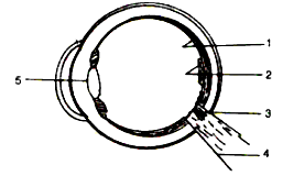
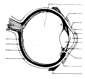
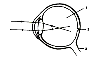
Given alongside is a diagram depicting a defect of the human eye. Study the same and then answer the questions that follow:
(i) Identify the defect.
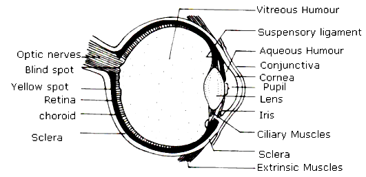
(ii) Name the parts labelled 1, 2 and 3.
(iii) Give labelled two possible reasons for this eye defect.
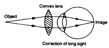
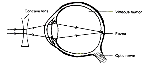
(iv) Draw a labelled diagram to show how the above mentioned defect is rectified.
(i) Identify the defect.
(ii) Name the parts labelled 1, 2 and 3.
(iii) Give labelled two possible reasons for this eye defect.
(iv) Draw a labelled diagram to show how the above mentioned defect is rectified.
Answer
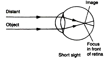
View full question & answer→(i) Myopia.
(ii) 1. Vitreous humor
2. Fovea
3. Optic nerve
(iii) 1. Lengthening of the eyeball from front to back.
2. Lens is too curved.
(iv) Correction of Myopia:
(ii) 1. Vitreous humor
2. Fovea
3. Optic nerve
(iii) 1. Lengthening of the eyeball from front to back.
2. Lens is too curved.
(iv) Correction of Myopia: