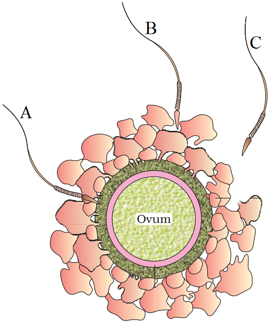
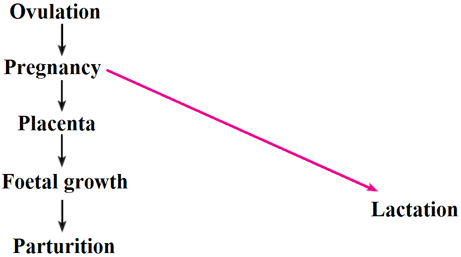
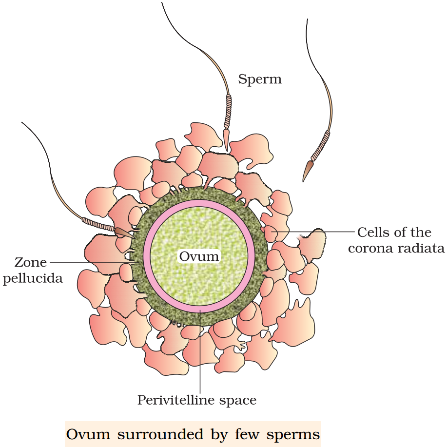
Month 1: Human development starts when a sperm fuses with an egg to create a zygote. A zygote is a single-cell that contains two copies of chromosomes-one copy from each parent. In the week following fertilization, the zygote undergoes rapid cell division and becomes a mass of cells known as a blastocyst. After more cell division, the blastocyst splits in half.
Month 2: This month, the embryo’s development shifts into high gear. Its tongue, teeth and eyelids start to form. Its limbs grow longer and stronger, and its palate is nearly complete. Also in this time period, the embryo’s gastrointestinal tract separates from its urogenital organs and its heart begins beating-twice as fast as yours, in fact.
Month 3: This is the month of the heartbeat. Using a special tool called a Doppler monitor, doctors can detect the tiny thump-thumps of a 10-week-old fetus.
Month 4: Get out the headphones and tune the radio dial to Mozart-this month, the fetus can hear its mother’s heartbeat, her voice and other outside noises. The fetus is also developing at warp speed; by now, all its major organs are complete.
Month 5: Finally, the baby really starts kicking! “Quickening” is when a mother senses her potential punter in action for the first time, and this milestone moment usually happens during pregnancy’s fifth month.
Month 6: Month 6 marks the end of the second trimester. At this point, the fetus’s essential organs-its kidneys, heart and lungs-are fully formed. The facial features are becoming more recognizable. It also can hiccup and react to loud “outside” noises by blinking, shifting or kicking. By month’s end, the fetus will measure about 12 or 13 inches long and weigh roughly 2 pounds.
Month 7: The 7-month-old fetus can blink, and its eyes may even remain open for short period of time. Hands and feet are becoming even more active. Also in this phase: Taste buds form and protective fat tissue makes its debut. The fetus’s hearing is fully developed and, in boys, its testicles have moved to the groin. By month’s end, the baby-to-be will measure 14 to 16 inches long and weigh anywhere from 2 ½ to 3 ½ pounds.
Month 8: This month, the fetus’s brain develops rapidly, and all of its organs except the lungs are mature. An 8-month-old fetus stretches 16 to 18 inches long and weighs between 4 and 6 pounds. And as the baby-to-be grows larger, space in the womb becomes scarce. Expectant mothers should still count on catching a few elbows every day, but the elaborate somersault sequences should stop until delivery day. Other exciting changes during this period? The fetus’s fingernails now reach beyond its fingertips and its skin is starting to smooth.
Month 9: The finish line is in sight! In this final month of development, the fetus’s fat layers thicken to help keep it warm outside the womb, and the protective layers of vernix caseosa and lanugo largely disappear. By now, the fetus’s lungs are mature, its skin pink and smooth, and its toenails fully grown. The baby-to-be can also execute an array of reflexes, such as head turning, blinking and grasping. At this late stage, it stretches between 20 to 22 inches long, and weighs about 7½ pounds.

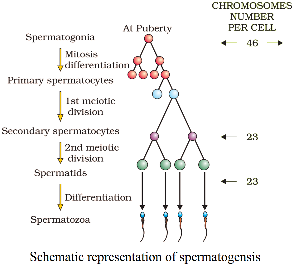
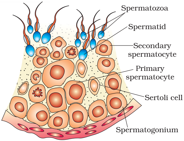 Diagrammatic sectional view of a seminiferous tubule (enlarged)Hormonal control of spermatogenesis.
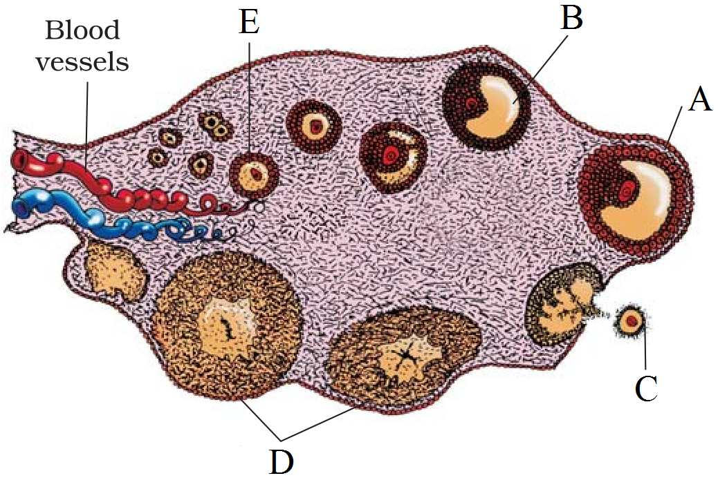
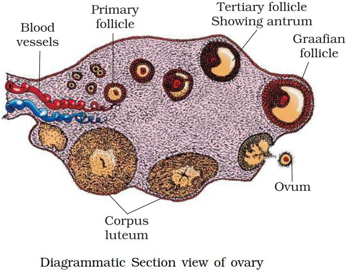
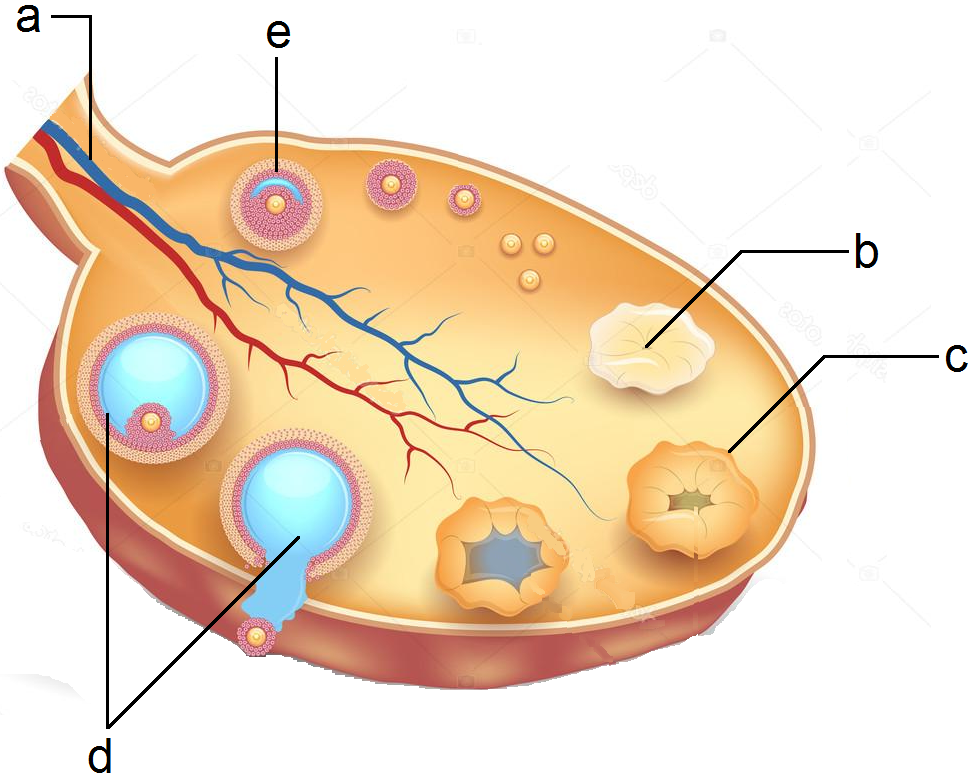
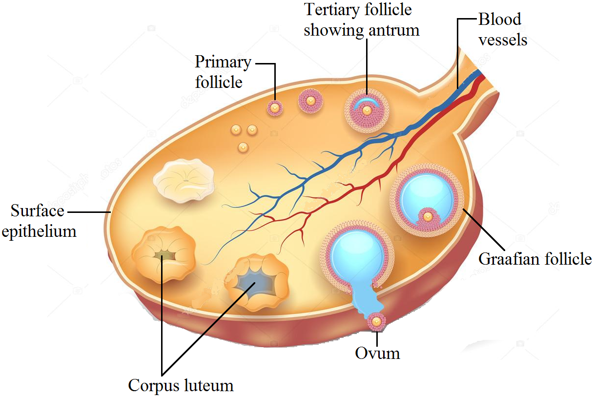
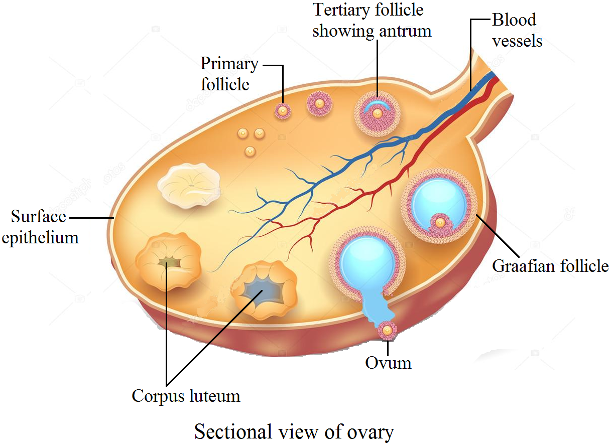 Sectional view of ovary.
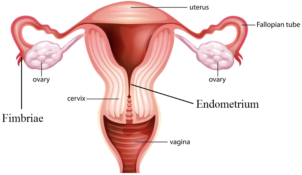
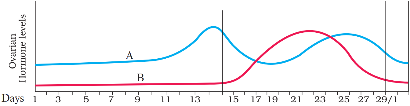
 Diagrammatic sectional view of a seminiferous tubule (enlarged) . Hormonal control of spermatogenesis.
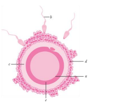
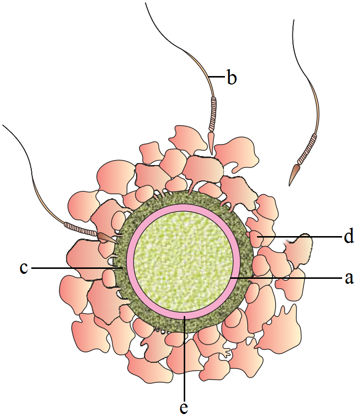
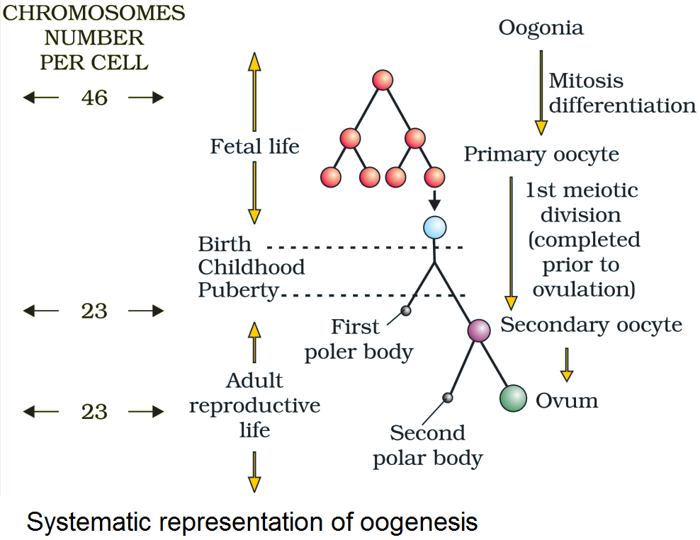
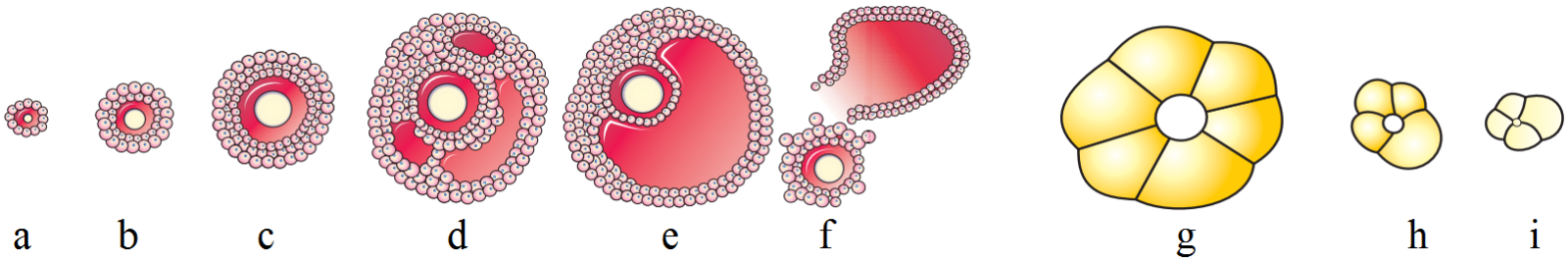
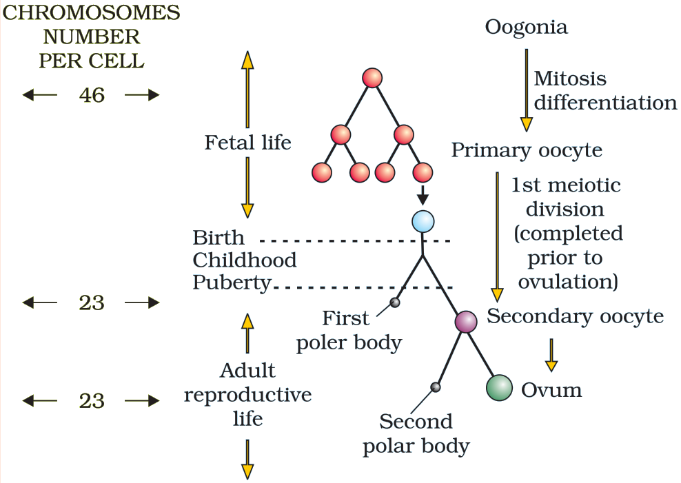
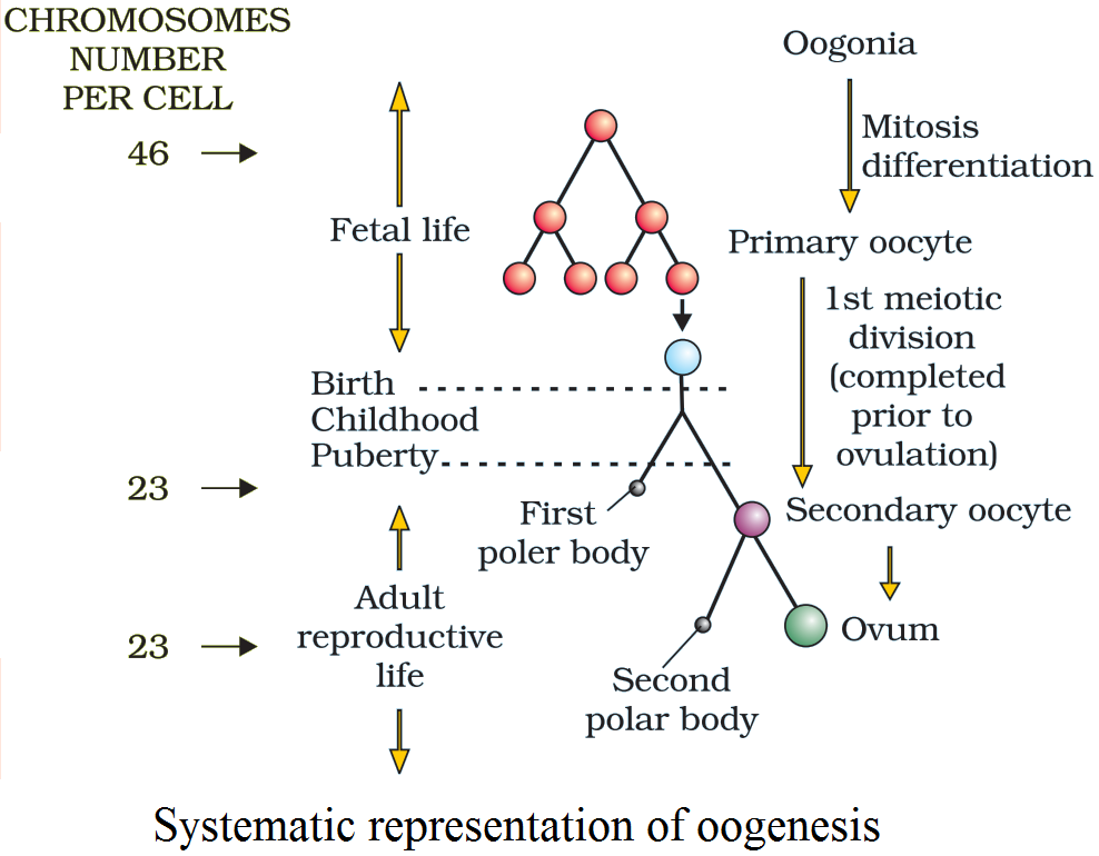 Systematic representation of oogenesis.
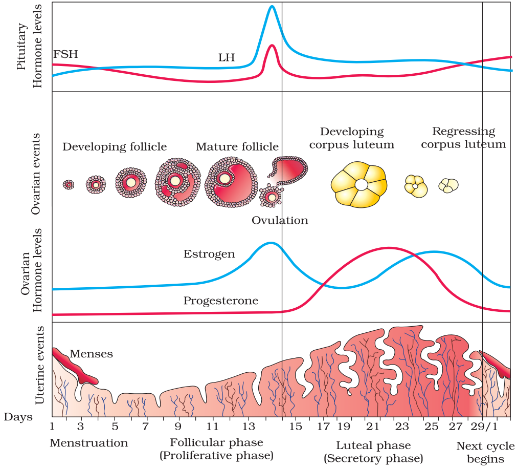
 Hypothalamus secretes gonadotropin releasing hormone (GnRH) which stimulates the anterior lobe of pituitary gland to secrete LH (Luteinising hormone) and FSH (Follicle stimulating hormone).