Questions

Case study (4 Marks)

Take a timed test

9 questions · self-marked practice — reveal the answer and mark yourself.

Question 14 Marks
Describe the process of translation during protein synthesis
Answer
→ Prokaryotic Translation involves following step.
(i) Activation of amino acid
(ii) Formation of polypeptide chain
→ Activation of amino acid
→ In the presence of aminoacyl tRNA Synthatare (Enzyme), a specific amino acid reacts with ATP
→ Amino acid + ATP + Enzyme $\xrightarrow{ Mg ^{2+}}$ Amino acid amp Enzyme (complex) + PPi
→ In aminoacylation of tRNA, the a.acid AMP - E (complex) reacts with uncharged t-RNA.
→ Amino acid - AMP (complex) - Enzyme + t-RNA → Amino acid (Charged) - t-RNA + Enzyme + PPi
→ Formation of Polypeptide chain
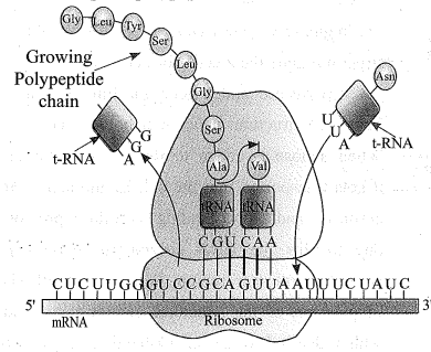
→ The cellular factory responsible for synthesising proteins is the ribosome.
→ The ribosome consists of structural RNAs and about 80 different proteins.
→ In its inactive state, it exists as two subunits; a large subunit and a small subunit.
→ When the small subunit encounters an mRNA, the process of translation of the mRNA to protein begins.
→ There are two sites in the large subunit, for subsequent amino acids to bind to and thus, be close enough to each other for the formation of a peptide bond.
→ The ribosome also acts as a catalyst (23S rRNA in bacteria is the enzyme - ribozyme) for the formation of peptide bond.
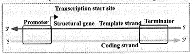
→ A translational unit in mRNA is the sequence of RNA that is flanked by the start codon (AUG) and the stop codon and codes for a
polypeptide.
→ An mRNA also has some additional sequences that are not traslated and the referred as untranslated regions (UTR). The UTRs are present at both 5'-end (before start codon) and at 3'- end (after stop codon). They arerequired for efficient translation process
→ Translation has 3 steps
• Initiation
→ For initiation, the ribosome binds to the mRNA at the start codon (AUG) that is recognised only by the initiator tRNA.
• Elongation
→ The ribosome proceeds to the elongation phase of protein synthesis. During this stage, complexes composed of an amino acid linked to tRNA, sequentially bind to the appropriate codon in mRNA by forming complementary base pairs with the tRNA anticodon.
→ The ribosome moves from codon to codon along the mRNA.
→ Amino acids are added one by one, translated into Polypeptide sequences dictated by DNA and represented by mRNA.
• Termination
→ At the end, a release factor binds to the stop codon, termination translation and releasing the complete polypeptide from the ribosome.
Image
View full question & answer
Question 24 Marks
Explain packaging of DNA Helix###Explain the formation of nucleosome in Eukaryotic cell
Answer
→ Taken the distance between two consecutive base pairs as 0.34 nm (0.34 x 10⁻⁹ m), if the length of DNA double helix in a typical mammalian cell is calculated (simply by multiplying the total number of bp with distance between two consecutive bp,
→ The length of DNA =Total no. of base pair x Distance between two base pair
Image
→ A length that is far, greater than the dimension 6 of a typical nucleus (approximately 10-6 m).
→ In prokaryotes, such as, E. coli, though they do not have a defined nucleus, the DNA is not scattered throughout the cell.
→ DNA (being negatively charged) is held with some proteins (that have positive charges) in a region termed as 'nucleoid'.
→ The DNA in nucleoid is organised in large loops held by proteins.
Image
→ In eukaryotes, this organisation is much more complex. There is a set of positively charged, basic proteins called histones.
→ A protein acquires charge depending upon the abundance of amino acids residues with charged side chains.
→ Histones are rich in the basic amino acid residues lysine and arginine. rek
→ Both the amino acid residues carry positive charges in their side chains.
→ Histones are organised to form a unit of eight molecules called histone octamer.
→ Nucleosome:
→ The negatively charged DNA is wrapped around the positively charged histone octamer to form a structure called nucleosome.
→ A typical nucleosome contains 200 bp of DNA helix.
→ Nucleosomes constitute the repeating unit of a structure in nucleus called chromatin, thread- like stained (coloured) bodies seen in nucleus.
→ The nucleosomes in chromatin are seen as 'beads-on-string' structure when viewed under electron microscope (EM).
→ The beads-on-string structure in chromatin is packaged to form chromatin fibers that are further coiled and condensed at metaphase stage of cell division to form chromosomes.
→ Non-histone Chromosomal (NHC) proteins:
→ The packaging of chromatin at higher level requires additional set of proteins that collectively are referred to as Non-histone Chromosomal (NHC) proteins.
→ Euchromatin :
→ In a typical nucleus, some region of chromatin are loosely packed (and stains light) and are referred to as euchromatin.
→ Heterochromatin:
→ The chromatin that is more densely packed and stains dark are known as heterochromatin. Euchromatin is said to be transcriptionally active chromatin, whereas heterochromatin is inactive.
View full question & answer
Question 34 Marks
Explain with diagram - Hershey and Chase experiment.###On the basis of experimental proof explain the diagram that DNA is genetic material.###How did Hershey and Chase differentiate between DNA and protein in their experiment while proving that DNA is the genetic material?
Answer
→ The unequivocal proof that DNA is the genetic material came from the experiments of Alfred Hershey and Martha Chase (1952). They worked with viruses that infect bacteria called bacteriophages.
→ The bacteriophage attaches to the bacteria and its genetic material then enters the bacterial cell.
→ The bacterial cell treats the viral genetic material as if it was its own and subsequently manufactures more virus particles.
→ Hershey and Chase worked to discover whether it was protein or DNA from the viruses that entered the bacteria.
→ They grew some viruses on a medium that contained radioactive phosphorus and some others on medium that contained radioactive sulfur.
→ Viruses grown in the presence of radioactive phosphorus contained radioactive DNA but not radioactive protein because DNA contains phosphorus but protein does not.
→ Similarly, viruses grown on radioactive sulfur contained radioactive protein but not radioactive DNA because DNA does not contain sulfur.
→ Radioactive phages were allowed to attach to E.coli bacteria.
→ Then, as the infection proceeded, the viral coats were removed from the bacteria by agitating them in a blender.
→ The virus particles were separated from the bacteria by spinning them in a centrifuge.
→ Bacteria which was infected with viruses that had radioactive DNA were radioactive, indicating that DNA was the material that passed from the virus to the bacteria.
→ Bacteria that were infected with viruses that had radioactive proteins were not radioactive.
→ This indicates that proteins did not enter the bacteria from the viruses.
→ DNA is therefore the genetic material that is passed from virus to bacteria.
Image
View full question & answer
Question 44 Marks
Write a short note on transcription unit.### Explain : Transcription unit
Answer
→ A transcription unit in DNA is defined primarily by the three regions in the DNA :
(i) The Structural gene
(ii) A Promoter
(iii) A Terminator
(i) A structural gene:
→ There is a convention in defining the two strands of the DNA in the structural gene of a transcription unit.
→ Since the two strands have opposite polarity and the DNA-dependent RNA polymerase also catalyse the polymerisation in only one direction, that is, 5' → 3',
→ Template strand:
→ The strand that has the polarity 3' → 5' acts as a template, and is also referred to as template strand.
Coding strand :
→ The other strand which has the polarity (5'→ 3') and the sequence same as RNA (except thymine at the place of uracil), is displaced during transcription. Strangely, this strand (which does not code for anything) is referred to as coding strand.
→ All the reference point while defining a transcription unit is made with coding strand.
Image
Schematic structure of a transcription unit
(ii) A promoter:
→ The promoter and terminator flank the structural gene in a transcription unit.
→ The promoter is said to be located towards 5'-end (upstream) of the structural gene (the reference is made with respect to the polarity of coding strand).
→ It is a DNA sequence that provides binding site for RNA polymerase and it is the presence of a promoter in a transcription unit that also defines the template and coding strands.
(iii) A Terminator:
→ By switching its position with terminator, the definition of coding and template strands could be reversed.
→ The terminator is located towards 3'-end (downstream) of the coding strand and it usually defines the end of the process of transcription.
→ There are additional regulatory sequences that may be present further upstream or downstream to the promoter.
View full question & answer
Question 54 Marks
Explain Methodology of HGP
Answer
→ The methods involved two major approaches. One approach focused on identifying all the genes that are expressed as RNA (referred to as Expressed Sequence Tags (ESTS).
→ The other took the blind approach of simply sequencing the whole set of genome that contained all the coding and non-coding sequence, and later assigning different regions in the sequence with functions (a term referred to as Sequence Annotation).
→ For sequencing, the total DNA from a cell is isolated and converted into random fragments of relatively smaller sizes (recall DNA is a very long polymer, and there are technical limitations in sequencing very long pieces of DNA) and cloned in suitable host using specialised vectors.
→ The cloning resulted into amplification of each piece of DNA fragment so that it subsequently could be sequenced with ease.
→ The commonly used hosts were bacteria and yeast, and the vectors were known as BAC (bacterial artificial chromosomes); and YAC (yeast artificial chromosomes).
→ The fragments were sequenced using automated DNA sequencers that worked on the principle of a method developed by Frederick Sanger. (Remember, Sanger is also credited for developing method for determination of amino acid sequences in proteins).
→ There sequences were then arranged based on some overlapping regions present in them.
→ This required generation of overlapping fragments for sequencing. Alignment of these sequences was humanly not possible.
→ Therefore, specialised computer based programs were developed.
→ These sequences were subsequently annotated and were assigned to each chromosome.
→ The sequence of chromosome 1 was completed only in May 2006 (this was the last of the 24 human chromosomes - 22 autosomes and X and Y - to be sequenced).
→ Another challenging task was assigning the genetic and physical maps on the genome.
→ This was generated using information on polymorphism of restriction endonuclease recognition sites, and some repetitive DNA sequences known as microsatellites (one of the applications of polymorphism in repetitive DNA sequences shall be explained in next section of DNA fingerprinting).
View full question & answer
Question 64 Marks
Explain different types of RNA along with process of transcription.
Answer
→ Type of RNA In bacteria, there are three major types of RNAs:
(i) mRNA (messenger RNA),
(ii) tRNA (transfer RNA), and
(iii) rRNA (ribosomal RNA).
→ All three RNAs are needed to synthesise a protein in a cell.
→ The mRNA provides the template, tRNA brings aminoacids and reads the genetic code, and rRNAs play structural and catalytic role during translation.
→ There is a single DNA-dependent RNA polymerase that catalyses transcription of all types of RNA in bacteria.
Process of Transciption
→ RNA polymerase binds to promoter and initiates transcription (Initiation).
It uses nucleoside triphosphates as substrate and polymerises in a template depended fashion following the rule of complementarity.
→ It somehow also facilitates opening of the helix and continues elongation.
→ Only a short stretch of RNA remains bound to the enzyme.
→ Once the polymerases reaches the terminator region, the nascent RNA falls off, so also the RNA polymerase. This results in termination of transcription.
→ An intriguing question is that how is the RNA polymerases able to catalyse all the three steps, which are initiation, elongation and termination.
→ The RNA polymerase is only capable of catalysing the process of elongation. It associates transiently with initiation-factor (σ) and termination-factor (p) to initiate and terminate the transcription, respectively.
→ Association with these factors alter the specificity of the RNA polymerase to either initiate or terminate.
Image
→ In bacteria, since the mRNA does not require any processing to become active, and also since transcription and translation take place in the same compartment (there is no separation of cytosol and nucleus in bacteria), many times the translation can begin much before the mRNA is fully transcribed.
→ Consequently, the transcription and translation can be coupled in bacteria.
View full question & answer
Question 74 Marks
Write a note on Mechanism of DNA replication
Answer
→ In living cells, such as E.coli, the process of replication requires a set of catalysts (enzymes).
→ The main enzyme is referred to as DNA- dependent DNA polymerase, since it uses a DNA template to catalyse the polymerisation of deoxynucleotides.
→ These enzymes are highly efficient as they have to catalyse polymerisation of a large number of nucleotides in a very short time.
→ E.coli that has only 4.6 x 10⁶ bp (compare it 6 with human whose diploid content is 6.6 x 10⁹ bp), completes the process of replication within 18 minutes; that means the average rate of polymerisation has to be approximately 2000 bp per second.
Not only do these polymerases have to be fast, but they also have to catalyse the reaction with high degree of accuracy.
Any mistake during replication would result into mutations. Furthermore, energetically replication is a very expensive process.
→ Deoxyribonucleoside triphosphates serve dual purposes.
(i) act as a substrates.
(ii) provide energy for polymerisation reaction (the two terminal phosphates in a deoxynucleoside triphosphates are high- energy phosphates, same as in case of ATP).
→ DNA-dependent DNA polymerases:
→ In addition to DNA-dependent DNA polymerases, many additional enzymes are required to complete the process of replication with high degree of accuracy.
→ Replication fork:
→ For long DNA molecules, since the two strands of DNA cannot be separated in its entire length (due to very high energy requirement), the replication occurs within a small opening of the DNA helix, referred to as replication fork.
→ Continuous, discontinuous. Strands:
→ The DNA-dependent DNA polymerases catalyse polymerisation only in one direction, that is 5' → 3'.
→ This creates some additional complications at the replicating fork.
→ Consequently, on one strand (the template with polarity 3' → 5'), the replication is continuous, while on the other (the template with polarity 5' → 3'), it is discontinuous.
→ The discontinuously synthesised fragments are later joined by the enzyme DNA ligase.
→ Origin of replication:
→ The DNA polymerases on their own cannot initiate the process of replication.
→ Also the replication does not initiate randomly at any place in DNA.
→ There is a definite region in E. coli DNA where the replication originates. Such regions are termed as origin of replication.
→ It is because of the requirement of the origin of replication that a piece of DNA if needed to be propagated during recombinant DNA procedures, requires a vector.
→ The vectors provide the origin of replication.
→ Cell division & DNA replication:
→ In eukaryotes, the replication of DNA takes place at S-phase of the cell-cycle.
→ The replication of DNA and cell division cycle should be highly coordinated.
→ A failure in cell division after DNA replication results into polyploidy (a chromosomal anomaly).
Image
View full question & answer
Question 84 Marks
Describe the process of DNA Fingerprinting in detail.
Answer
→ The technique of DNA Fingerprinting was initially developed by Alec Jeffreys. He used a satellite DNA as probe that shows very high degree of polymorphism.
→ It was known as Variable Number of Tendem Repeats (VNTR).
→ The technique, as used earlier, involved Southern blot hybridisation using radiolabelled VNTR as a probe. It included
(i) isolation of DNA,
(ii) digestion of DNA by restriction endonucleases,
(iii) seperation of DNA fragments by electrophoresis,
(iv) transferrring (blotting) of separated DNA fragments to synthetic membranes, such as nitrocellulose or nylon,
(v) hybridisation using labelled VNTR probe, and
(vi) detection of hybridised DNA fragmenents by autoradiography. A schematic representation of DNA fingerprinting is shown in Figure.
→ The VNTR belongs to a class of satellite DNA referred to as mini-satellite. A small DNA sequence is arranged tandemly in many copy numbers.
→ The copy number varies from chromosome to chromosome in an individual.
→ The numbers of repeat show very high degree of polymorphism. As a result the size of VNTR varies in size from 0.1 to 20 kb. Consequently, after hybridisation with VNTR probe, the autoradiogram gives many bands of differing sizes.
→ There bands give a characteristic pattern for an individual DNA.
→ It differs from individual to individual in a population except in the case of monozygotic (identical) twins.
→ The sensitivity of the technique has increased by use of polymerase chain reaction (PCR).
→ Consequently, DNA from a single cell is enough to perform DNA fingerprinting analysis. In addition to application in forensic science, it has much wider application, such as in determining population and genetic diversities.
→ Currently, many different probes are used to generate DNA fingerprints.
Image
View full question & answer
Question 94 Marks
Write a note Lac operon
Answer
→ The elucidation of the lac operon was also a result of a close association between a geneticist, Francois Jacob and a biochemist, Jacque Monod.
→ They were the first to elucidate a transcriptionally regulated system.
→ In lac operon (here lac refers to lactose), a polycistronic structural gene is regulated by a common promoter and regulatory genes.
→ Such arrangement is very common in bacteria and is referred to as operon.
→ The lac operon consists of one regulatory gene (the i gene here the term i does not refer to inducer, rather it is derived from the word inhibitor) and three structural genes (z, y, and a).
→ The i gene codes for the repressor of the lac operon.
→ The z gene codes for beta-galactosidase (β-gal), which is primarily responsible for the hydrolysis of the disaccharide, lactose into its monomeric units, galactose and glucose.
→ The y gene codes for permease, which increases permeability of the cell to ẞ-galactosides.
→ The a gene encodes a transacetylase.
→ Hence, all the three gene products in lac operon are required for metabolism of lactose. In most other operons as well, the genes present in the operon are needed together to function in the same or related metabolic pathway.
→ Inducer:
→ Lactose is the substrate for the enzyme beta- galactosidase and it regulates switching on and off of the operon. Hence, it is termed as inducer.
→ In the absence of a perferred carbon source such as glucose, if lactose is provided in the growth medium of the bacteria, the lactose is transported into the cells through the action of permease (Remember, a very low level of expression of lac operon has to be present in the cell all the time, otherwise lactose cannot enter the cells).
→ The lactose then induces the operon in the following manner.
→ The repressor of the operon is synthesised (all- the-time-constitutively) from the i gene. The repressor protein binds to the operator region of the operon and prevents RNA polymerase from transcribing the operon.
→ In the presence of an inducer, such as lactose or allolactose, the repressor is inactivated by interaction with the inducer.
→ This allows RNA polymerase access to the promoter and transcription proceeds. Essentially, regulation of lac operon can also be visualised as regulation of enzyme synthesis by its substrate.
→ Remember, glucose or galactose cannot act as inducers for lac operon.
→ Lac operon would remain expressed untill all the amount of lactose is converted to glucose & Galactose.
→ Regulation of lac operon by repressor is referred to as negative regulation. Lac operon is under control of positive regulation as well, but it is beyond the scope of discussion at this level.
Image
View full question & answer