Question 15 Marks
What is oogenesis? Give a brief account of oogenesis.
Answer
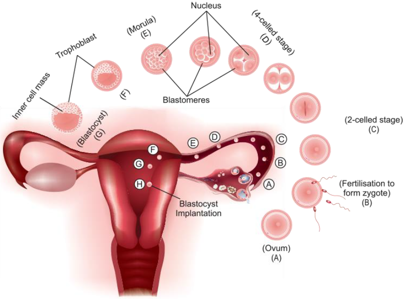
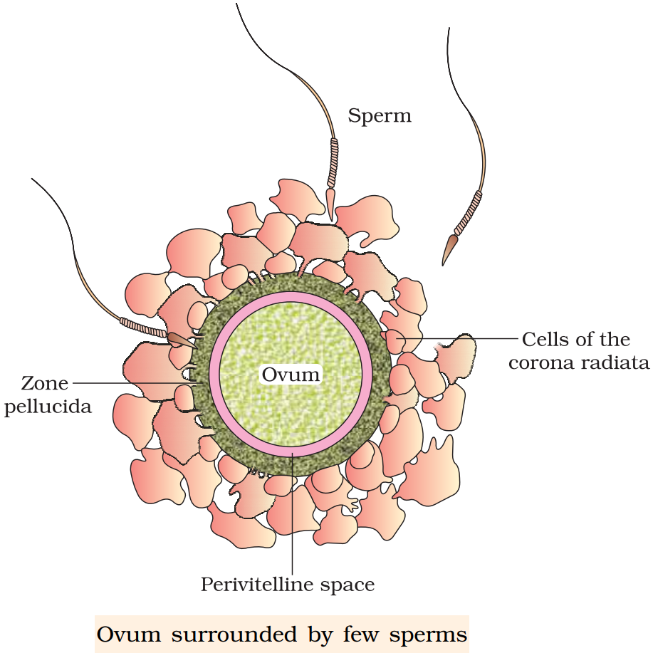
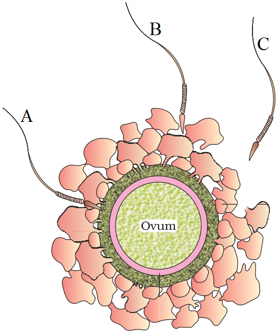
View full question & answer→The process of the formation of a mature ovum in the ovary from the oogonia in females is known as oogenesis.
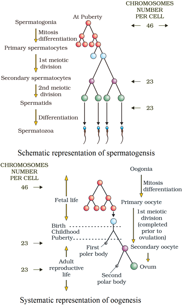
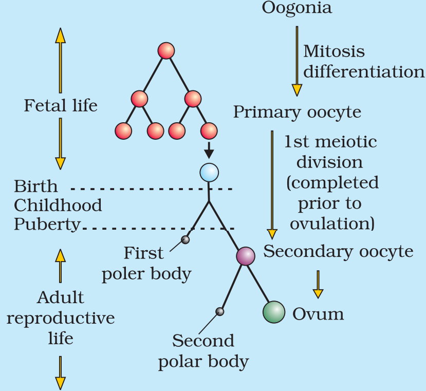
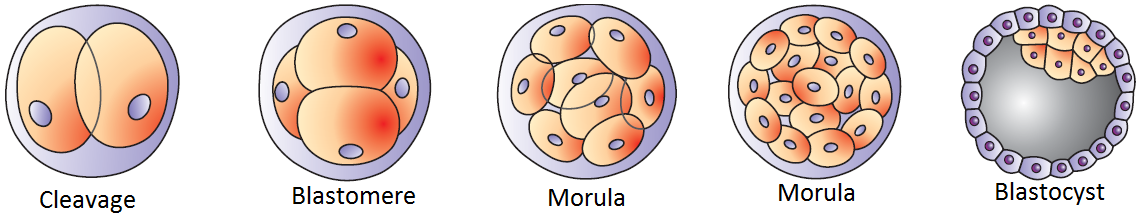
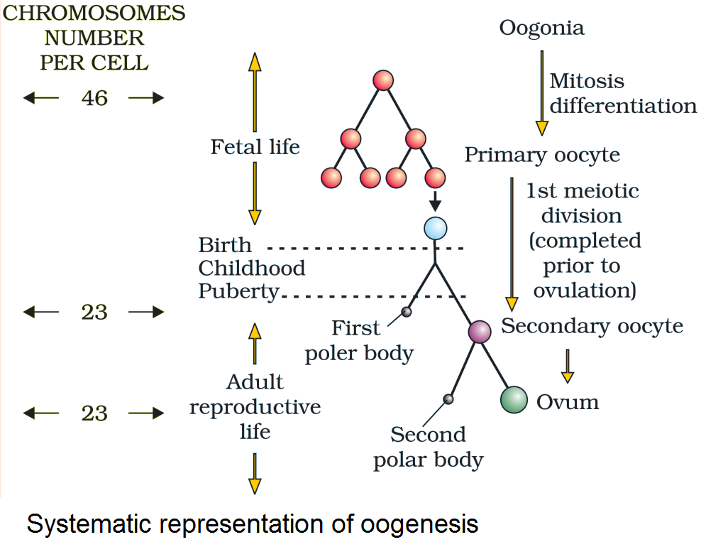
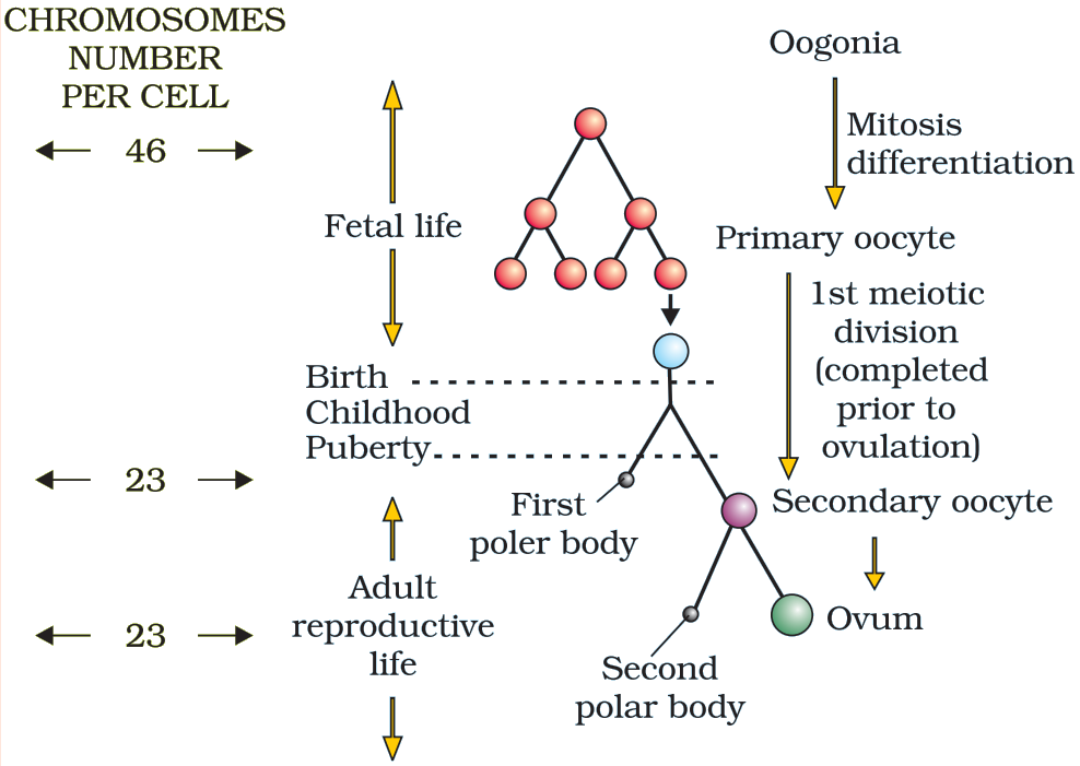
Germ cell of the female foetus divide to produce oogonia (gamete mother cell). No more oogonia are formed or added after birth. A diploid oogonium or egg mother cell increases in size and gets transformed into a diploid primary oocyte. This diploid primary oocyte undergoes meiosis or reductional division to form two unequal haploid cells. The smaller cell is known as the first polar body, while the larger cell is known as the secondary oocyte. This secondary oocyte undergoes meiosis II or equational division and gives rise to a second polar body and an ovum. Hence, in the process of oogenesis, a diploid oogonium produces a single haploid ovum while two or three polar bodies are produced.
Germ cell of the female foetus divide to produce oogonia (gamete mother cell). No more oogonia are formed or added after birth. A diploid oogonium or egg mother cell increases in size and gets transformed into a diploid primary oocyte. This diploid primary oocyte undergoes meiosis or reductional division to form two unequal haploid cells. The smaller cell is known as the first polar body, while the larger cell is known as the secondary oocyte. This secondary oocyte undergoes meiosis II or equational division and gives rise to a second polar body and an ovum. Hence, in the process of oogenesis, a diploid oogonium produces a single haploid ovum while two or three polar bodies are produced.

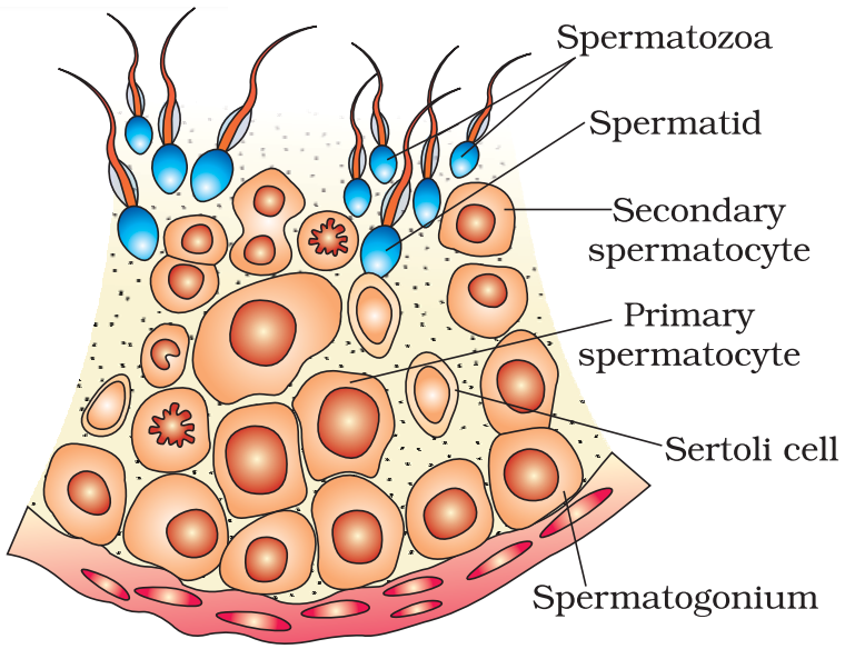 Diagrammatic sectional view of a seminiferous tubule (enlarged). Hormonal control of spermatogenesis.
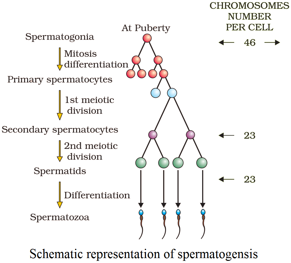 Schematic representation of spermatogensis.
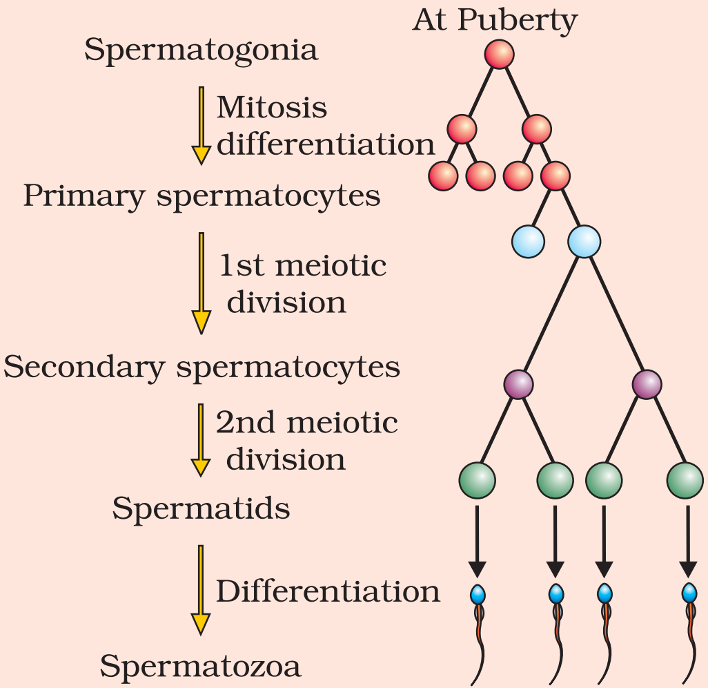
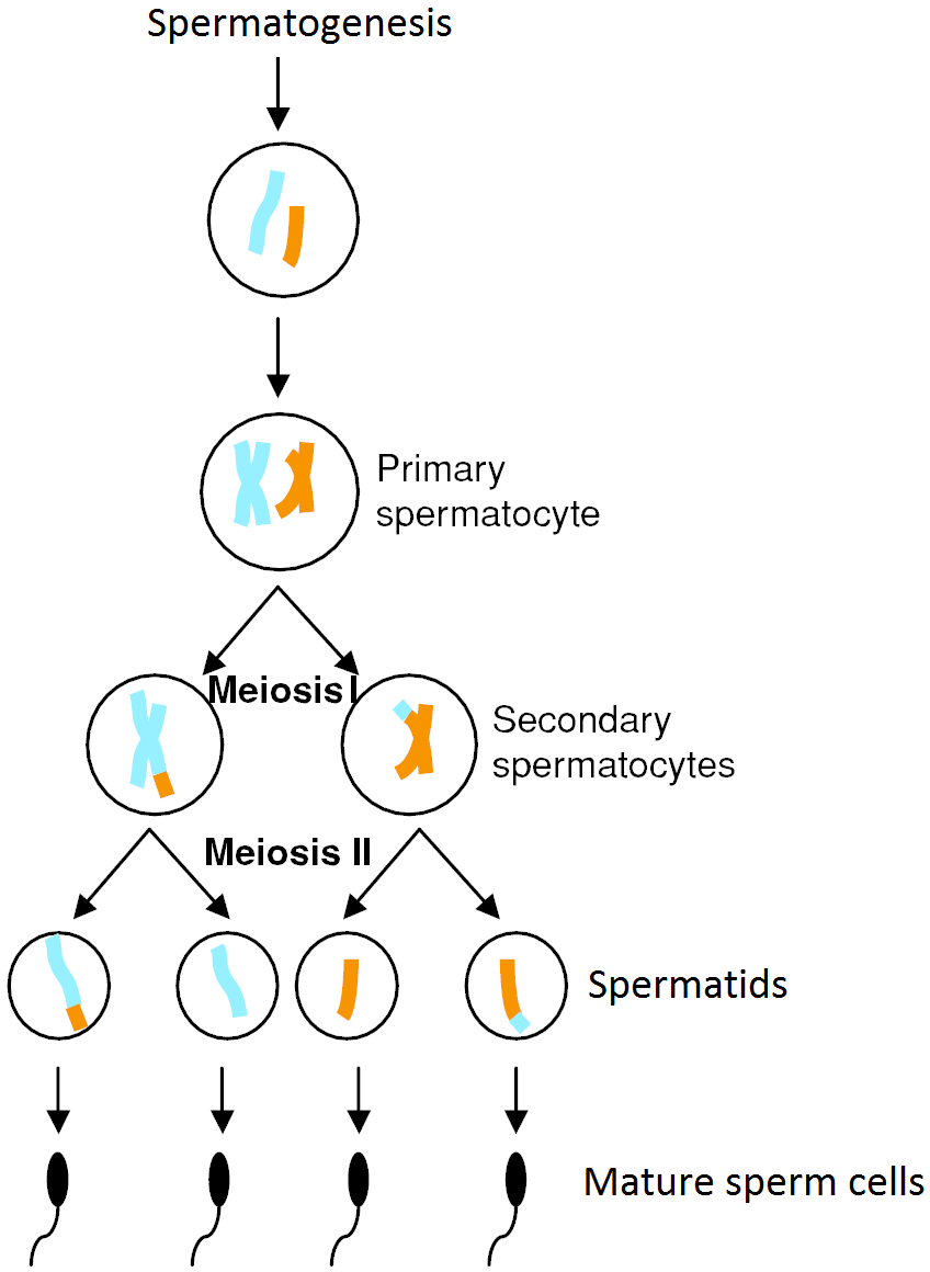
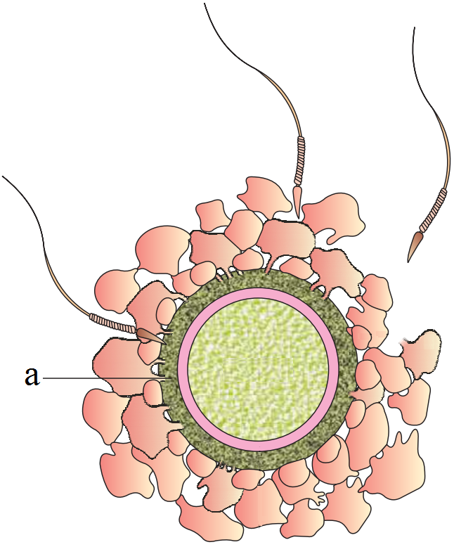


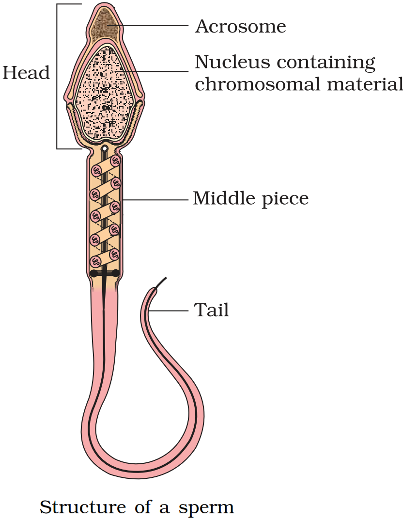
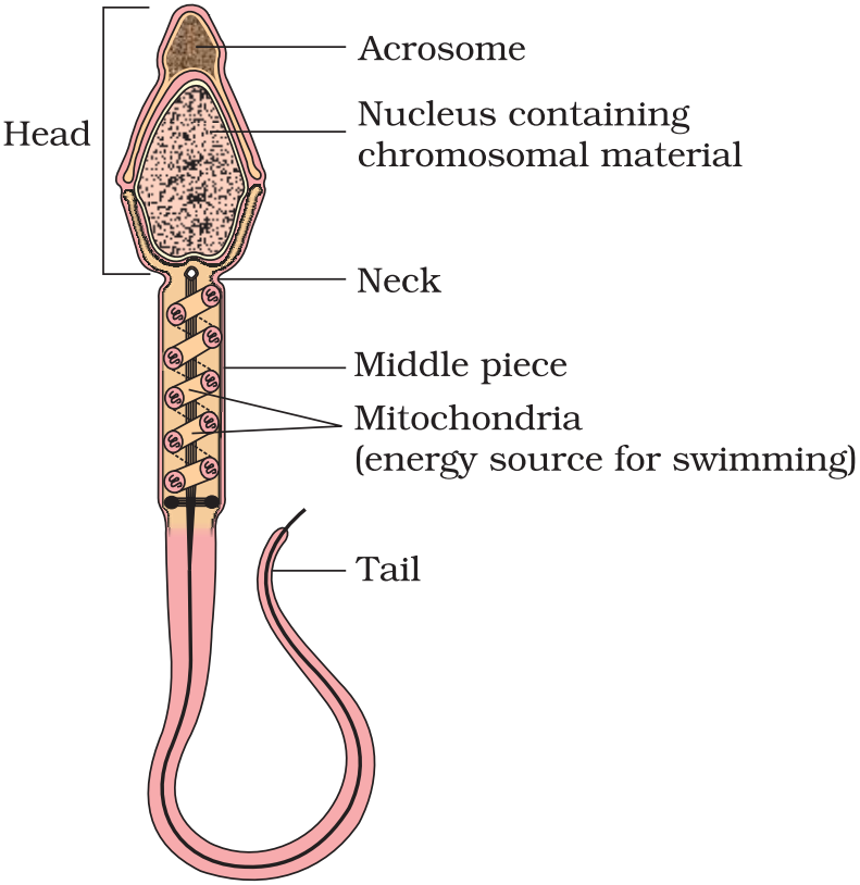 Structure of a sperm.
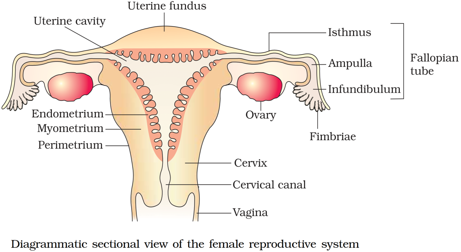


 Diagrammatic sectional view of a seminiferous tubule (enlarged)Hormonal control of spermatogenesis.
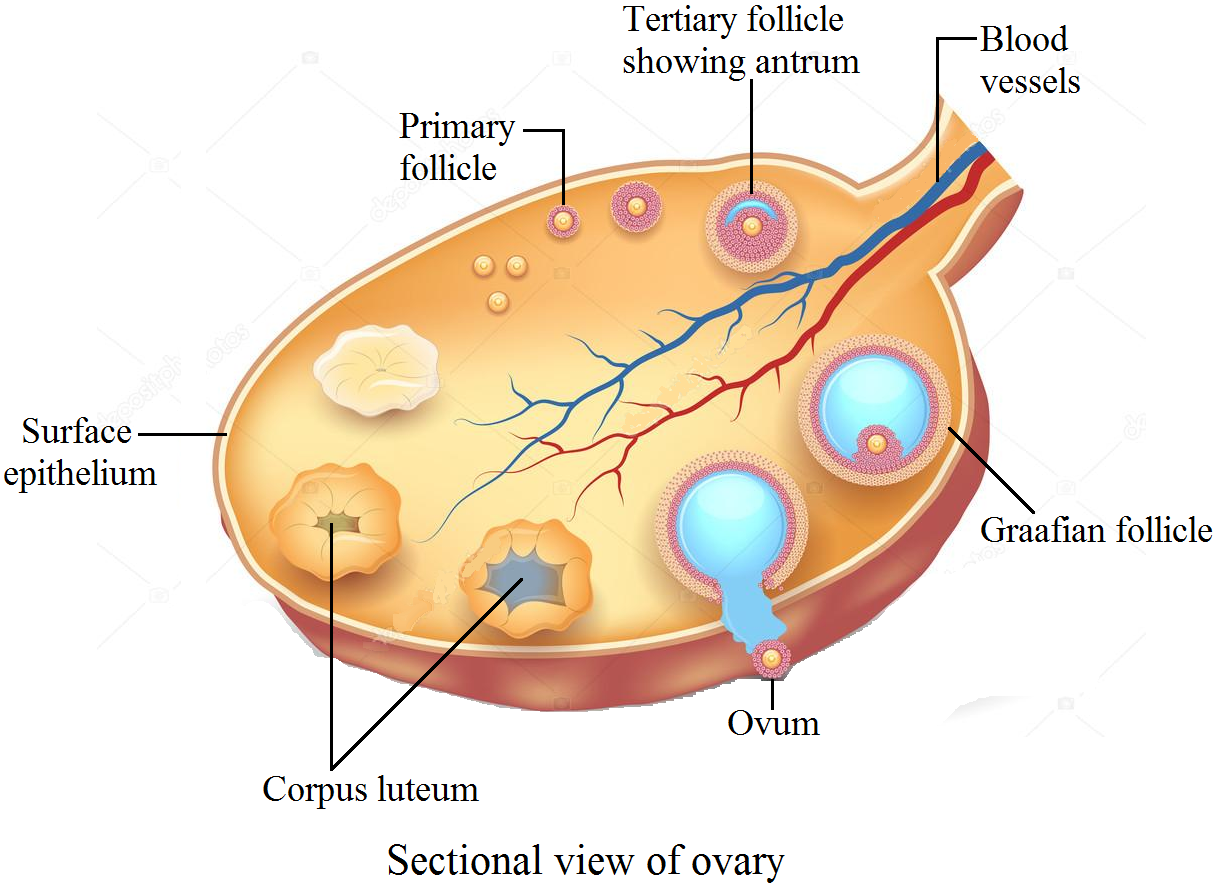 Sectional view of ovary.
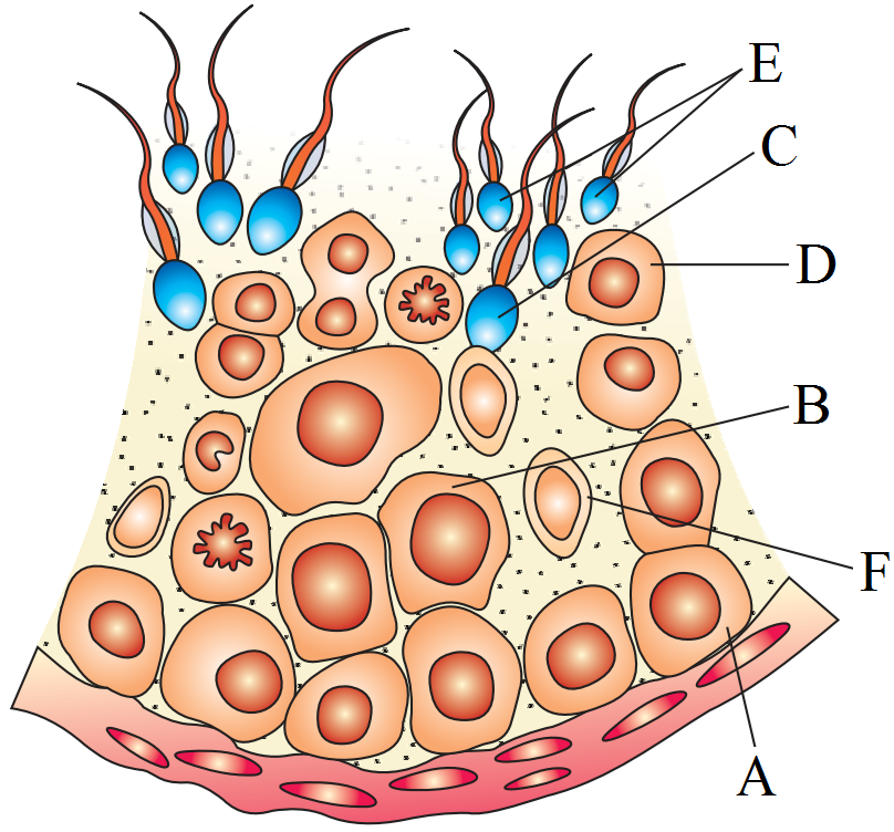
 Diagrammatic sectional view of a seminiferous tubule (enlarged) . Hormonal control of spermatogenesis.
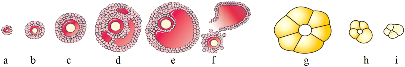
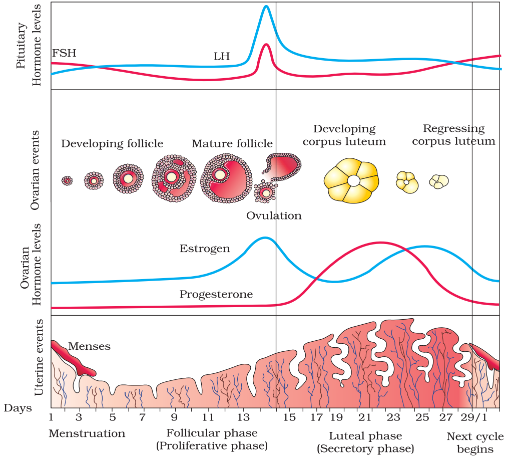
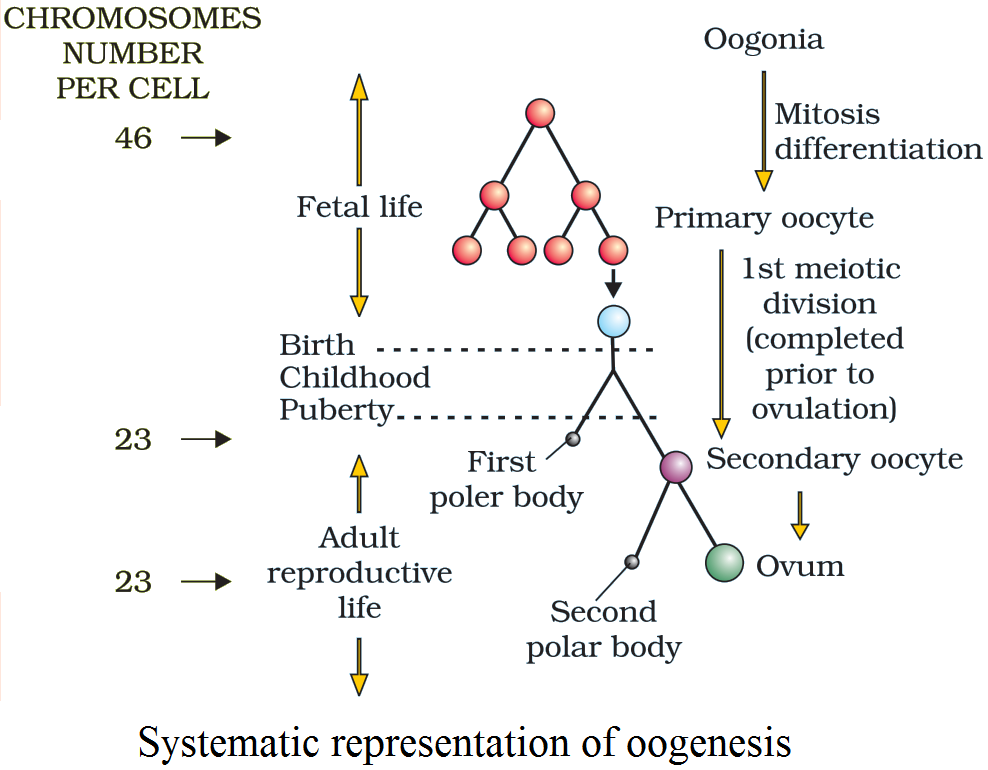 Systematic representation of oogenesis.
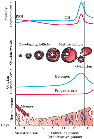
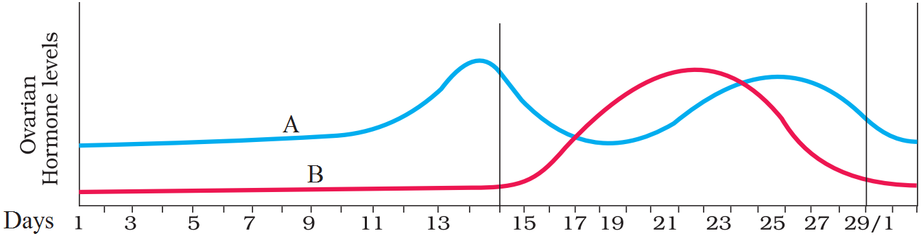
 Hypothalamus secretes gonadotropin releasing hormone (GnRH) which stimulates the anterior lobe of pituitary gland to secrete LH (Luteinising hormone) and FSH (Follicle stimulating hormone).
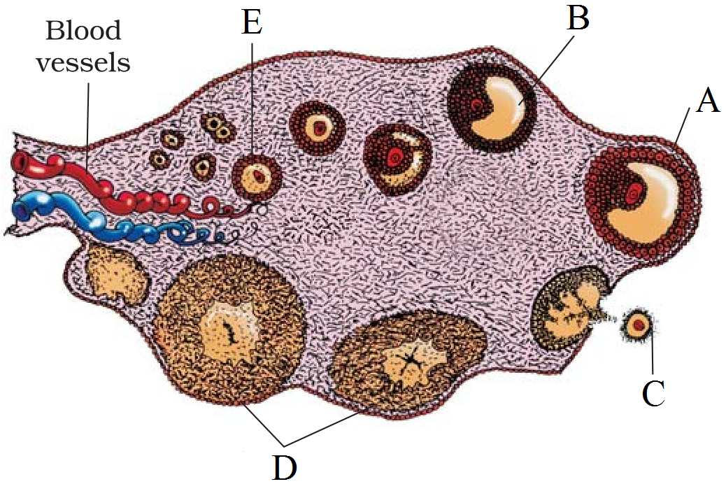
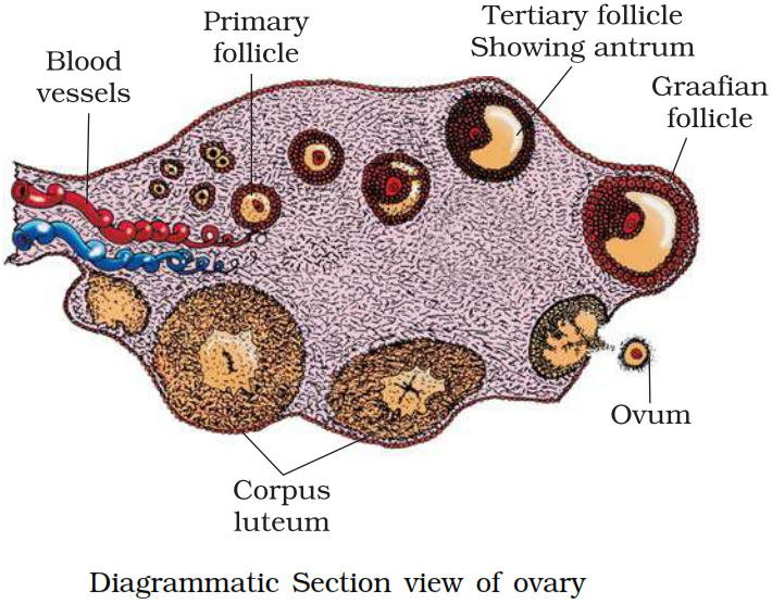


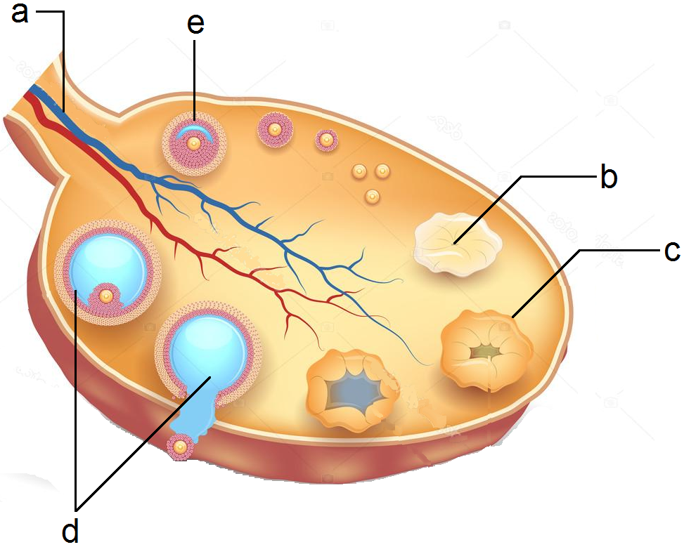


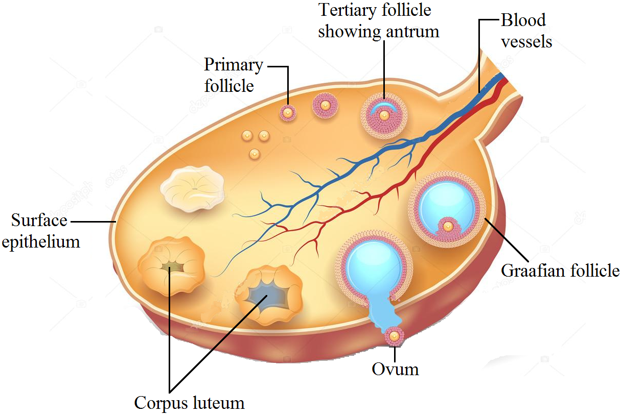

 Sectional view of ovary.


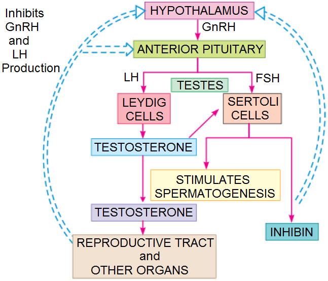 Flow chart showing the hormonal control of the human male reproductive system.