Epithelial tissue is the thin protective layer of cells which covers the surface of the body and lines the internal organs. The cells of this tissue are generally packed close together. The shape of the cells depends on the location and function of the tissue. Epithelial tissue originates from the ectoderm. But, epithelial tissue lining the intestine originates from the endoderm.
Epithelial tissue may be simple, i.e., composed of single layer of cells or stratified, i.e., made up of several layers of cells. Depending upon the shape and function of cells epithelial tissues are classified as:
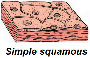
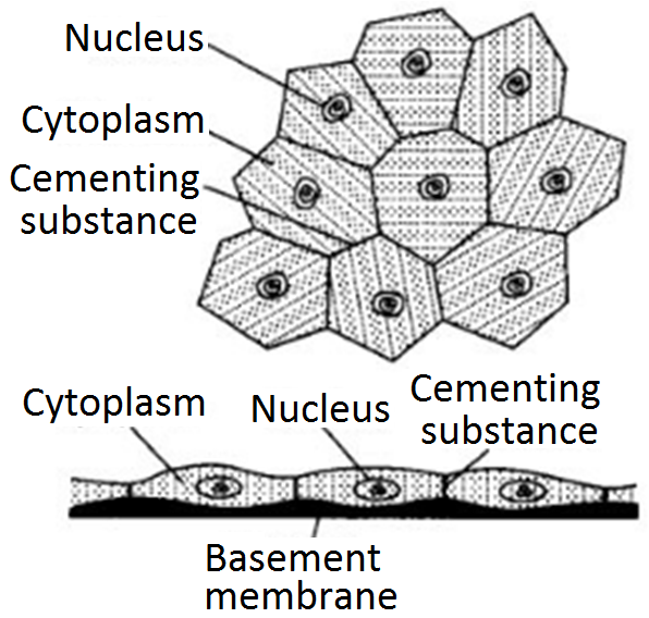
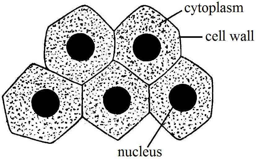
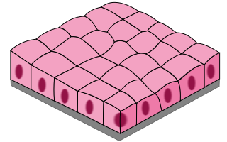
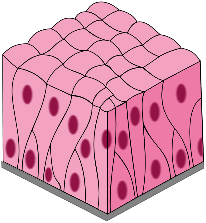
- Squamous epithelial tissue: This tissue is composed of a single layer of thin and flat, plate like ceils. The cells fit closely, like the bricks in a wall, to form a smooth membrane. It is also known as tesselated and pavement epithelium. It is found in the outer layer of the skin, and covers internal cavities and ducts. Tongue, oesophagus and the lining of the mouth are made up of squamous epithelium.It is also found in blood vessels and alveoli. It protects the underlying parts of body from mechanical injury, entry of germs, chemicals and drying. It also forms a selectively permeable surface through which filtration occurs.
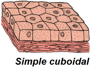
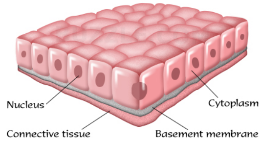
- Cuboidal epithelial tissue: This tissue is composed of cube like cells that fit closely. The cells look like squares in section, but the free surface appears hexagonal. This tissue lines the inside of the kidney tubules (the tubes leading from the cups of nephrons) thyroid vesicles and in glands like sweat glands, exocrine pancreas and the salivary glands. It forms germinal epithelium of gonads (testes and ovaries). It helps in absorption, excretion and secretion. It also provides mechanical support.
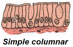
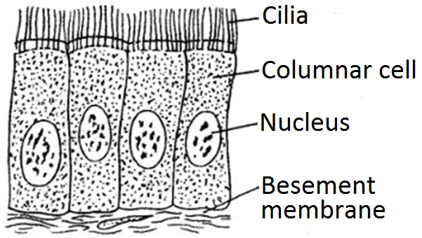
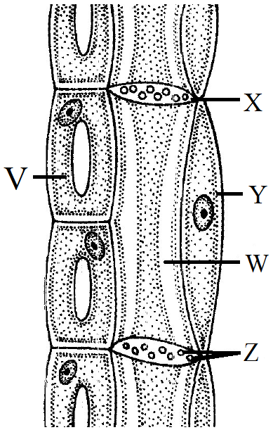
- Columnar ciliated epithelial tissue: This tissue is generally composed of a single layer of column like cells. The presence of a conspicuous striated border of microvilli at the free surface end of each cell increases the surface area of the cell for absorption and secretion. It is generally found in the inner lining of the alimentary canal. It also forms the lining of gall bladder and oviducts. The major functions of this tissue includes secretion (e.g., mucus of goblet cells) and absorption (e.g., stomach and intestine).
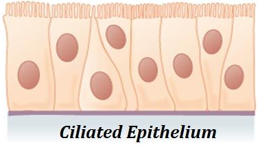
In some parts of the body, columnar epithelium develops protoplasmic outgrowths called cilia. The constant lashing movements of the cilia help to move substances. It is found in the sperm ducts. It also lines the trachea (wind-pipe), bronchi (lungs), kidney tubules and oviducts (Fallopian tubes). Ciliated epithelium helps the movement of ova in the fallopian tubes and the movement of mucus in the respiratory tract.
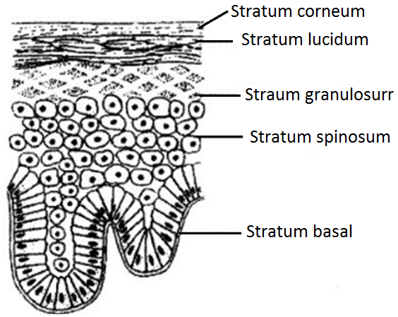
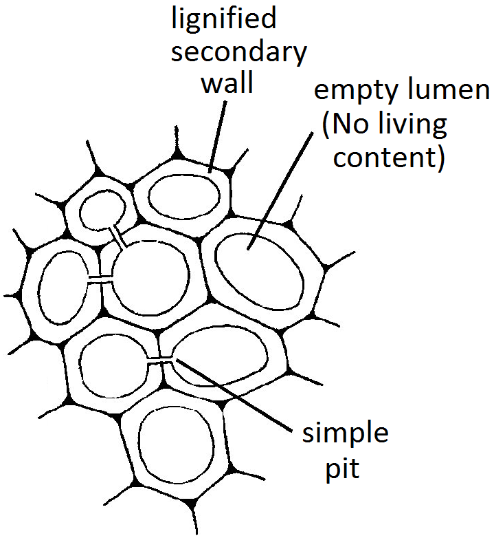
- Stratified squamous epithelial tissue: This tissue is found in skin and covers the external dry surface of the skin. Cells of this tissue are arranged in many layers, but the cells forming different layers of this epithelium are not similar. Deeper layers of the tissue have cuboidal cells which become polygonal and finally flattened (squamous) towards the free surface. The flattened cells of superficial layer may contain a fibrous protein, the keratin and become, dead cells and are called keratinised stratified squamous epithelium. This epithelium is water proof and highly resistant to mechanical injury.
- Glandular epithelial tissue: Epithelial tissue often acquire additional specialisation as gland cells, which can secrete substances at the epithelial surface. Sometimes, a portion of epithelial tissue folds in wards and a multicellular gland is formed. This is called glandular epithelium.
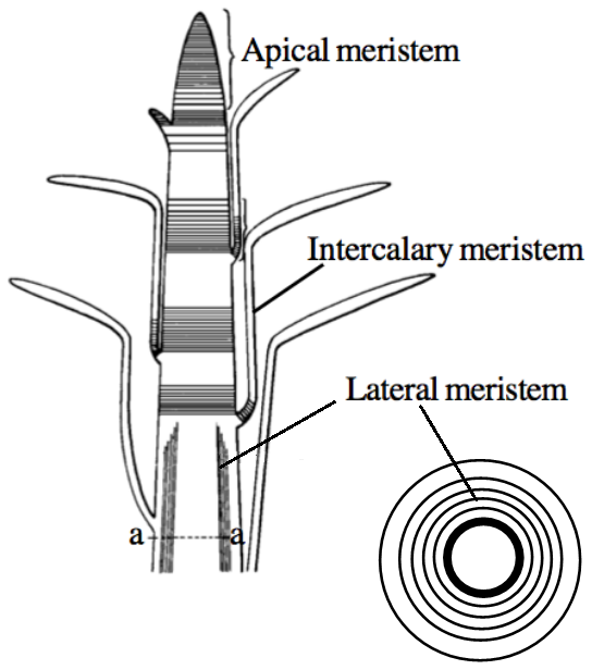
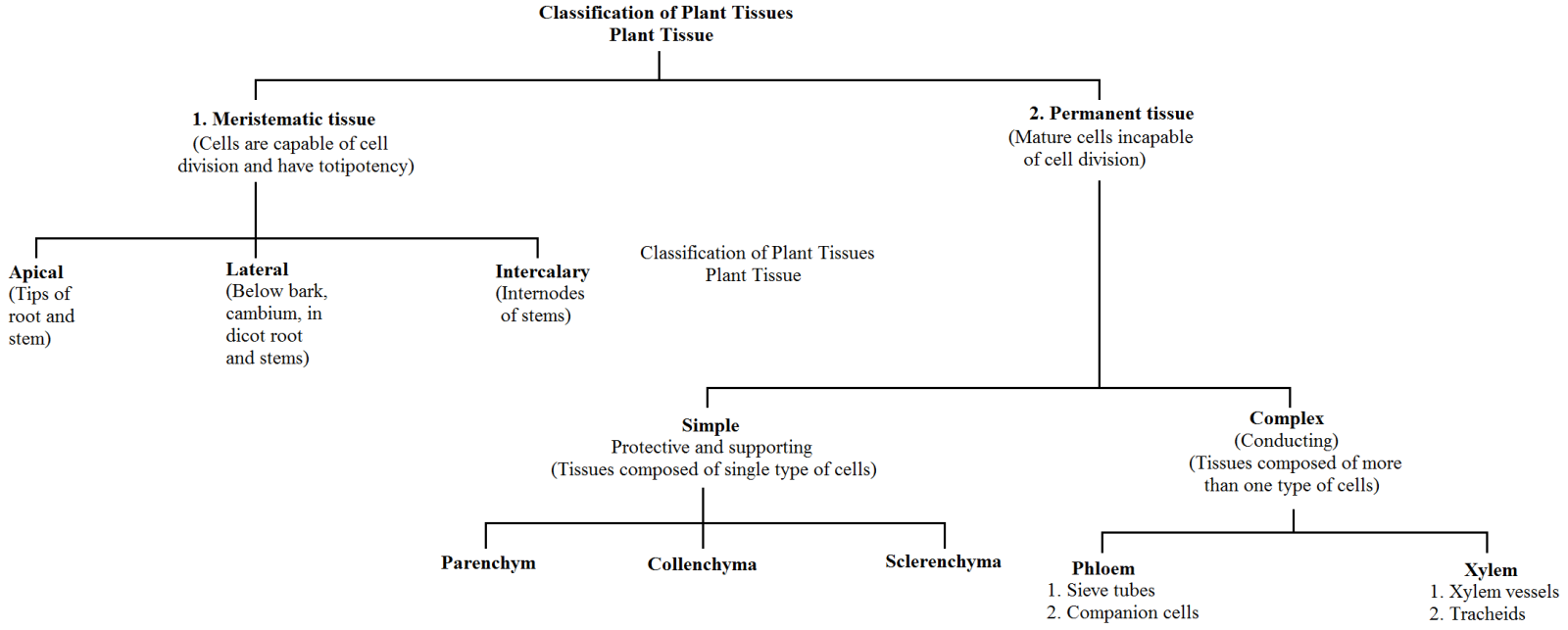
 According to their position in the plant, meristems are of three types:
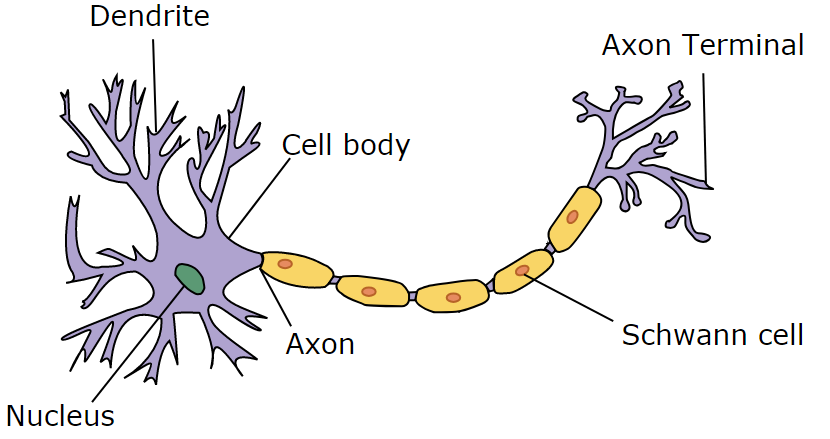
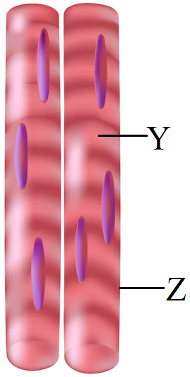
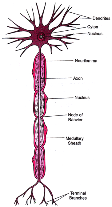
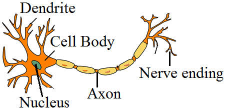 Cell body or cyton has a prominent nucleus and cytoplasm. Cell organelles like golgi bodies, mitochondria, etc are also present in the cytoplasm. From the cell body extend out two kinds of cytoplasmic extensions called dentrites and axons. The axon is covered by a fatty myelin sheath. Myelin sheath is discontinuous and broken at intervals by nodes of Ranvier. Axons usually a long, unbranched, cylindrical process that ends in many terminal end fibres. The axon ending of one nerve cell is lossely placed on the cell body of another nerve cell. The other small branch given out by the cyton are called dendrons which branch further into numerous thin hair-like dentrites.